3-dimensional cardiac outline reconstruction method

A contour reconstruction and heart technology, applied in medical science, image data processing, instruments, etc., can solve the problems of accurately generating three-dimensional heart phantom limitations, non-adaptive spatial filtering method, inaccurate signal source estimation, etc.

Active Publication Date: 2016-11-16
KOREA RES INST OF STANDARDS & SCI
View PDF2 Cites 3 Cited by
  • Summary
  • Abstract
  • Description
  • Claims
  • Application Information

AI Technical Summary

Problems solved by technology

[0005] However, non-adaptive spatial filtering methods may produce inaccurate estimates of deep signal sources, and thus, it may be limited in accurately generating 3D cardiac phantoms

Method used


Image

  • 3-dimensional cardiac outline reconstruction method
  • 3-dimensional cardiac outline reconstruction method
  • 3-dimensional cardiac outline reconstruction method

Examples


Embodiment Construction

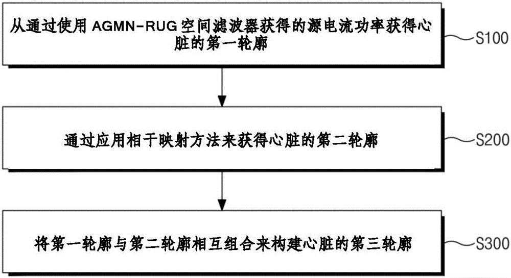
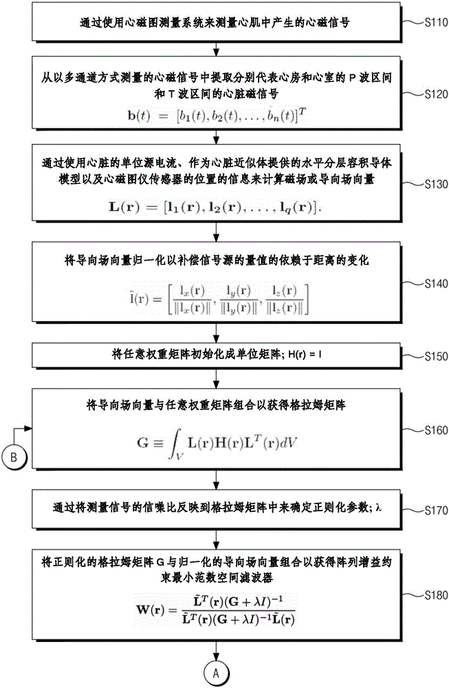
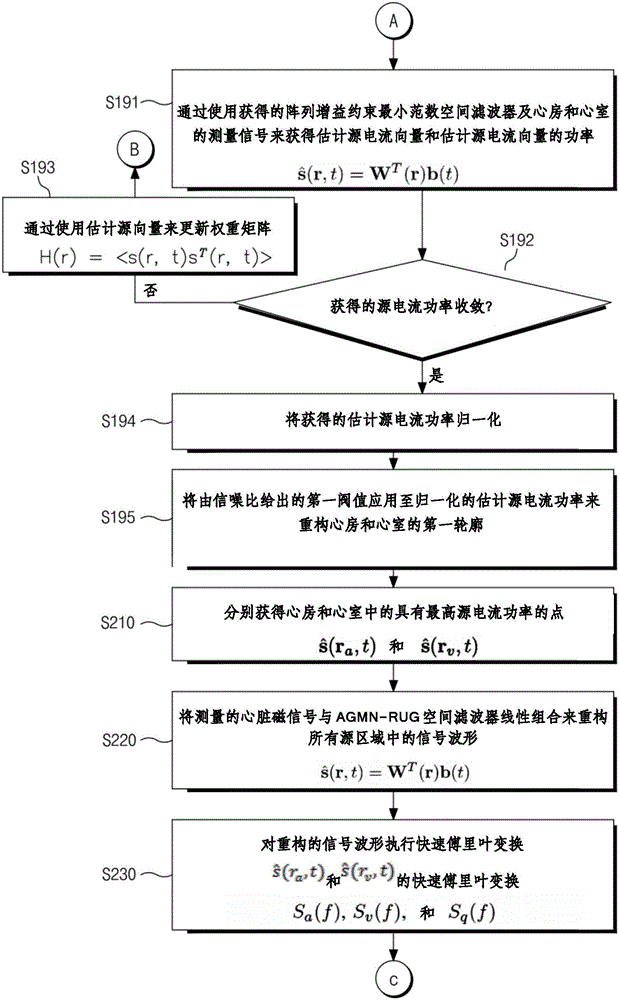
[0030]Three-dimensional cardiac visualization or mapping methods are useful for clinical applications of magnetocardiography (MCG). However, cardiac reconstruction requires additional image modalities. The present invention proposes a method for reconstructing a three-dimensional heart contour by using only MCG measurement data without using additional imaging techniques. Heart contours can be reconstructed by combining spatial filtering methods with coherence mapping methods.

[0031] The intensity of cardiac activity can be represented by an array-gain constraint minimum-norm with recursively-updated Gram matrix (AGMN-RUG) spatial filter.

[0032] Furthermore, the coherence between the maximum source point of the atrium and all source points can be compared with the coherence between the maximum source point of the ventricle and all source points by a coherence mapping method.

[0033] In numerical simulations and phantom experiments, the original shape can be compared wit...


PUM


Abstract

The present invention provides a cardiac outline reconstruction method and a magneto cardiogram measuring system. The cardiac outline reconstruction method comprises the steps of: obtaining a first cardiac outline from power of source current vector (source current power) obtained using an array-gain constraint minimum-norm with recursively updated gram matrix; AGMN-RUG) spatial filter; obtaining a second cardiac outline by applying a coherence mapping method; and configuring a third cardiac outline by coupling the first cardiac outline and the second cardiac outline with each other.

Description

technical field [0001] The present invention relates to a cardiac contour reconstruction method, and more particularly to a method of obtaining a three-dimensional cardiac contour from multi-channel data measured by a magnetocardiographic measurement system without the use of an anatomical imaging system such as an MRI system. Background technique [0002] A magnetocardiography (MCG) system is configured to measure the magnetic signals generated by the electrical activity of the myocardium by using a superconducting quantum interference device (SQUID) sensor as an ultrasensitive magnetic sensor, and non-invasively Diagnostic approach to heart disease. The location of cardiac disease can be localized by using multi-channel data measured by a magnetocardiographic measurement system. In particular, the localization of the locations of heart diseases in a three-dimensional heart phantom may allow doctors to visually observe information about the locations of disease loci. [0...

Claims


Application Information

Patent Timeline
Patent Type & Authority Applications(China)
IPC IPC(8): A61B5/00G06T7/00
CPCA61B5/243
Inventor 金基雄河泰勋权赫灿李龙镐金鎭穆
Owner KOREA RES INST OF STANDARDS & SCI
Why Eureka
Try Eureka