
Image blood vessel segmentation method and device

A blood vessel and image technology, applied in the field of medical images, can solve time-consuming problems, achieve clean blood vessel segmentation, and solve the effect of blood vessel segmentation problems

Active Publication Date: 2016-11-23
SHANGHAI UNITED IMAGING HEALTHCARE
View PDF8 Cites 29 Cited by
  • Summary
  • Abstract
  • Description
  • Claims
  • Application Information

AI Technical Summary

Problems solved by technology

This method needs to scan the patient twice, which is time-consuming and brings more scanning radiation to the patient

Method used


Image

  • Image blood vessel segmentation method and device
  • Image blood vessel segmentation method and device
  • Image blood vessel segmentation method and device

Examples


Embodiment Construction

[0019] In order to make the above objects, features and advantages of the present invention more comprehensible, specific implementations of the present invention will be described in detail below in conjunction with the accompanying drawings. In the following description, specific details are set forth in order to provide a thorough understanding of the present invention. However, the present invention can be implemented in many other ways than those described here, and those skilled in the art can make similar extensions without departing from the connotation of the present invention. Accordingly, the present invention is not limited to the specific embodiments disclosed below.

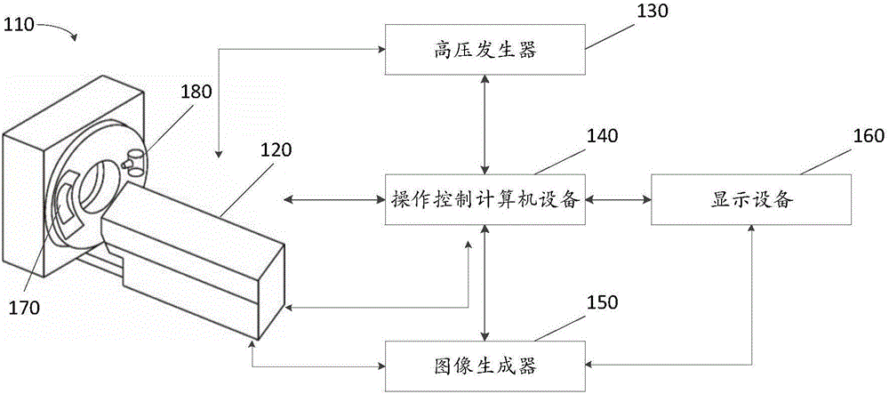
[0020] figure 1 Shown is a schematic diagram of an imaging system 100 . In some embodiments, the imaging system 100 can scan a predetermined target, obtain scan data and generate an image related thereto. In some embodiments, imaging system 100 may perform further processing on the generated imag...


PUM


Abstract

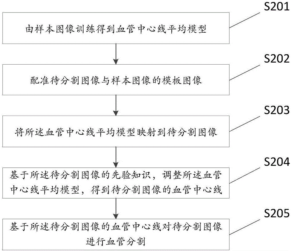
The invention discloses a blood vessel segmentation method and device. The method includes: using sample image training to obtain a blood vessel central line average model; aligning a to-be-segmented image with the template image of a sample image; mapping the blood vessel central line average model to the to-be-segmented image; regulating the blood vessel central line average model on the basis of the priori knowledge of the to-be-segmented image to obtain the blood vessel central line of the to-be-segmented image; performing blood vessel segmentation on the to-be-segmented image on the basis of the blood vessel central line of the to-be-segmented image. By the method, blood vessel segmentation problems can be well solved, blood vessel segmentation is clean, and segmentation failure caused by the fact that the bone is segmented.

Description

technical field [0001] The invention relates to the field of medical images, in particular to blood vessel segmentation. Background technique [0002] Vascular imaging techniques include computed tomography angiography (CTA), magnetic resonance angiography (MRA), etc., which are very important techniques that can help doctors diagnose various diseases of blood vessels, such as calcification, stenosis, aneurysm, etc. In the process of vascular imaging, three-dimensional images are mainly obtained, which not only show the blood vessels but also include other tissues around the blood vessels (such as fat, muscle, bone, etc.), which cannot give doctors an intuitive feeling. Therefore, it is very important to extract blood vessels from the overall 3D image and display the shape of blood vessels with 3D display technology for the diagnosis of vascular diseases. [0003] The techniques of CTA blood vessel extraction can be mainly divided into two categories: model method and non-m...

Claims


Application Information

Patent Timeline
Patent Type & Authority Applications(China)
IPC IPC(8): G06T7/00
CPCG06T2207/10072G06T2207/20081G06T2207/30101
Inventor 赵鹏
Owner SHANGHAI UNITED IMAGING HEALTHCARE
Learn More