Method for automatically identifying leukocytes in leucorrhea microscopic image

A microscopic image and automatic recognition technology, applied in the field of medical digital image processing, can solve problems such as high work intensity, easy to pollute the environment, unfavorable clinical diagnosis, etc.

Active Publication Date: 2017-01-04
UNIV OF ELECTRONICS SCI & TECH OF CHINA
View PDF3 Cites 36 Cited by
  • Summary
  • Abstract
  • Description
  • Claims
  • Application Information

AI Technical Summary

Problems solved by technology

Most of the traditional microscopic cell detection is manual microscopic examination. This method takes a long time to operate, has high work intensity, is easy to pollute the environment, and is subject to subjective influence, which is not conducive to clinical diagnosis.

Method used


Image

  • Method for automatically identifying leukocytes in leucorrhea microscopic image
  • Method for automatically identifying leukocytes in leucorrhea microscopic image
  • Method for automatically identifying leukocytes in leucorrhea microscopic image

Examples


Embodiment Construction

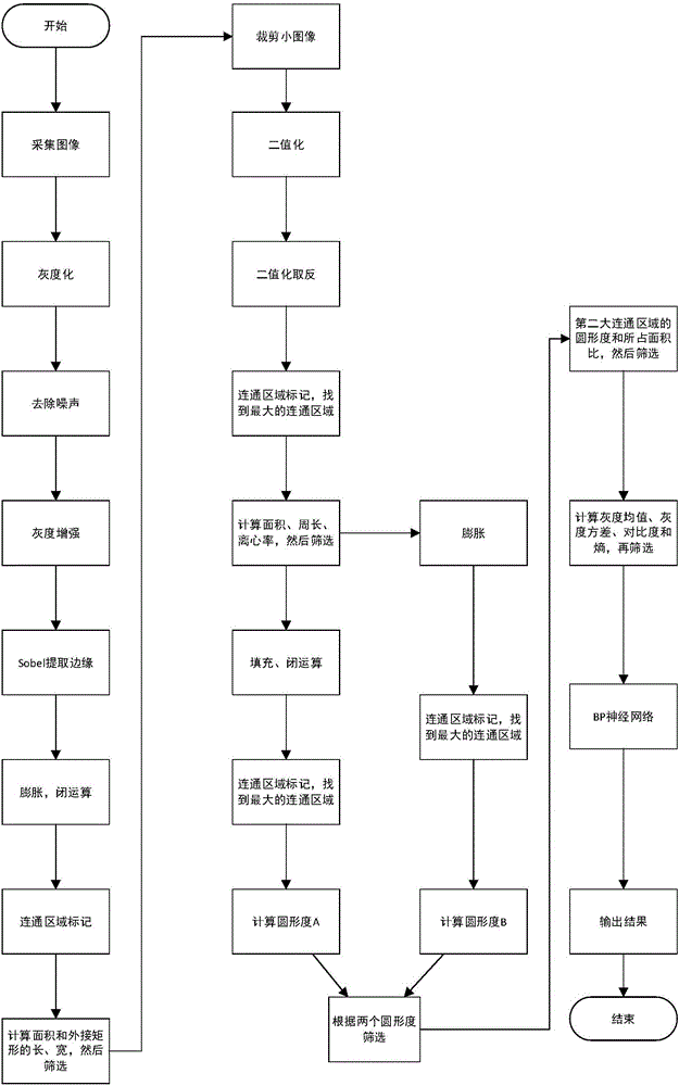
[0074] Below in conjunction with accompanying drawing, the automatic identification algorithm of a kind of leucorrhea white blood cell of the present invention is described in detail:

[0075] Step 1: Use a microscopic imaging system to automatically collect images of the formed components of cells;

[0076] Step 2: Perform grayscale processing on the image to obtain a grayscale image;

[0077] Step 3: Denoising the grayscale image obtained in step 2 by using a median filter to obtain a denoised image;

[0078] Step 4: using the method of histogram equalization on the image obtained in step 3 to enhance the contrast of the image to obtain a grayscale enhanced image;

[0079] Step 5: using the Sobel operator to extract the edge from the image obtained in step 4 to obtain an edge image;

[0080] Step 6: expand the image obtained in step 5 to obtain an expanded image;

[0081] Step 7: Perform a closed operation on the image obtained in step 6 to obtain an image after the close...


PUM


Abstract

The invention discloses a method for automatically identifying leukocytes in a leucorrhea microscopic image, and belongs to the field of medical digital image processing, particularly an algorithm for automatically identifying leukocytes in a leucorrhea microscopic image. According to the method, the microscopic image is subjected to gray-scale processing, then communication regions in the image are searched, the communication regions in the image are sequentially screened according to actual morphologies of the leukocytes, and finally, a leukocyte image in leucorrhea is identified, so that working time of a worker and personal errors are greatly reduced, and working efficiency is improved.

Description

technical field [0001] The invention belongs to the field of medical digital image processing, and specifically refers to an automatic identification algorithm for white blood cells in a leucorrhea microscopic image. Background technique [0002] Leucorrhea is the secretion of female vagina, and the detection of white blood cells in leucorrhea is an important condition for judging whether the female reproductive system is healthy. Most of the traditional microscopic cell detection is manual microscopic examination. This method takes a long time to operate, has high work intensity, is easy to pollute the environment, and is subject to subjective influence, which is not conducive to clinical diagnosis. In recent years, with the development of artificial intelligence research and the improvement of biomedical image processing technology, it has become a reality to use image processing technology to automatically identify the formed components of microscopic cell images. In the...

Claims


Application Information

Patent Timeline
Patent Type & Authority Applications(China)
IPC IPC(8): G06K9/00G06N3/08G06T5/00G06T7/00G06T7/60
CPCG06N3/084G06T5/002G06T5/009G06T7/60G06T2207/20032G06T2207/10061G06V20/695G06V20/698
Inventor 张静胡静蓉郝如茜王强张正龙刘娟秀倪光明杜晓辉刘霖刘永
Owner UNIV OF ELECTRONICS SCI & TECH OF CHINA
Why Eureka
Try Eureka