Surgery image processing method and surgery image processing device

An image processing and surgery technology, applied in the field of medical devices, can solve the problems of high lesion recognition ability, high labor cost, and low recognition efficiency of doctors, and achieve the goal of improving image recognition efficiency, reducing labor cost, and strengthening lesion recognition ability Effect

Inactive Publication Date: 2017-01-11
厚凯(天津)医疗科技有限公司
View PDF10 Cites 7 Cited by
  • Summary
  • Abstract
  • Description
  • Claims
  • Application Information

AI Technical Summary

Problems solved by technology

[0003] In the existing surgical image processing method, the doctor manually recognizes the lesion tissue information contained in the surgical image, the recognition efficiency is low, and the doctor's lesion recognition ability is high, and only experienced doctors can perform lesion tissue recognition
Therefore, the existing surgical image processing methods have low recognition efficiency and high labor costs.

Method used


Image

  • Surgery image processing method and surgery image processing device
  • Surgery image processing method and surgery image processing device
  • Surgery image processing method and surgery image processing device

Examples


Embodiment 1

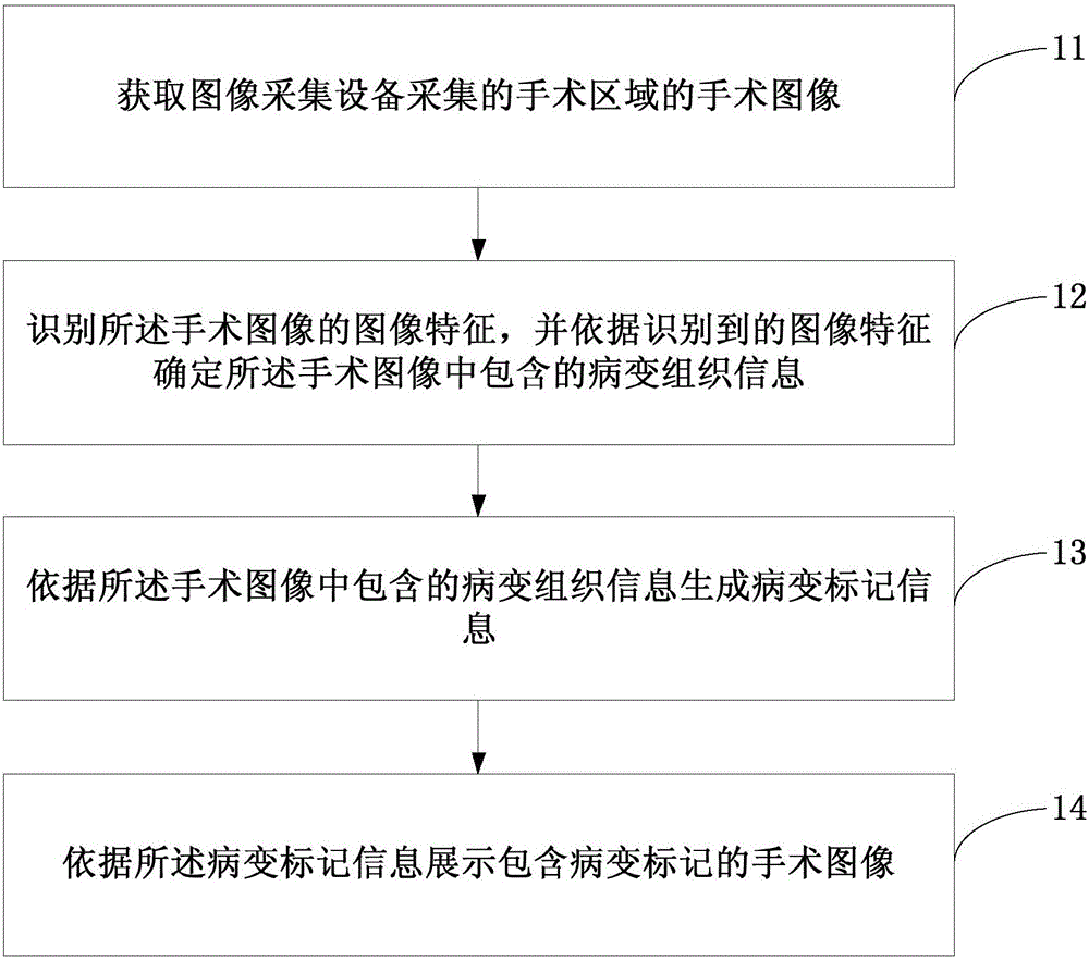
[0023] figure 1 It is a schematic flowchart of a surgical image processing method provided in Embodiment 1 of the present invention. The method can be executed by a surgical image processing device, where the device can be implemented by software and / or hardware. Such as figure 1 As shown, the implementation process includes:

[0024] Step 11. Obtain the operation image of the operation area collected by the image acquisition device.

[0025] In this embodiment, the image acquisition device is used to acquire patient images during ultrasonic surgery, for example, the image acquisition device may include endoscopes and / or microscopes and other devices. Specifically, in an ultrasonic surgery, an image acquisition device such as an endoscope and / or a microscope is aimed at an operation area of ​​a patient to obtain an operation image of the operation area.

[0026] Step 12, identifying image features of the surgical image, and determining lesion tissue information contained in...

Embodiment 2

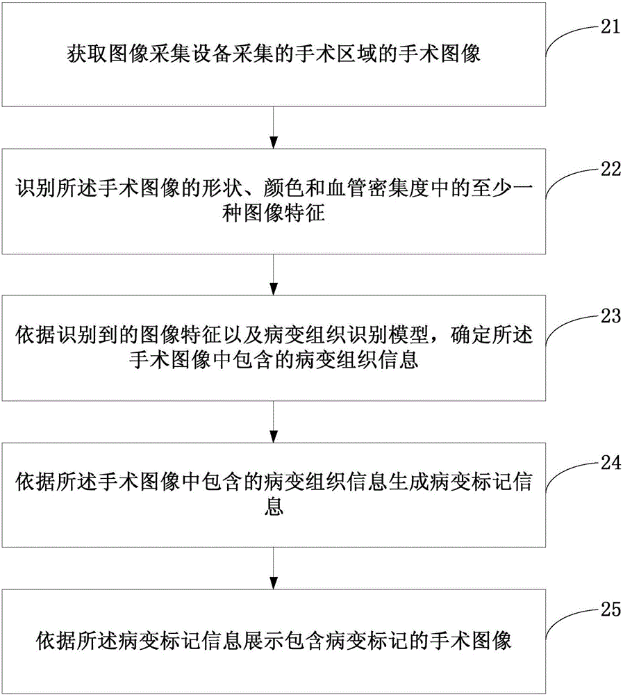
[0038] This embodiment provides a new surgical image processing method on the basis of the first embodiment above. image 3 A schematic flow chart of the surgical image processing method provided in Embodiment 2 of the present invention, as shown in image 3 As shown, the implementation process includes:

[0039] Step 21. Obtain the operation image of the operation area collected by the image acquisition device.

[0040] In this embodiment, the image acquisition device may be an image acquisition device used in surgical operations such as an endoscope or a microscope.

[0041] Step 22, identifying at least one image feature among shape, color, and blood vessel density of the surgical image.

[0042] Specifically, image recognition technology can be used for basic processing of surgical images to obtain different regions included in the surgical image, and then image features such as shape, color, or blood vessel density of different regions can be obtained.

[0043] Step 23...

Embodiment 3

[0051] Figure 4 It is a schematic structural diagram of the surgical image processing device provided by the third embodiment of the present invention. Such as Figure 4As shown, the specific structure of the surgical image processing device may include:

[0052] An image acquisition unit 31, configured to acquire an operation image of the operation area collected by the image acquisition device;

[0053] A lesion identifying unit 32, configured to identify image features of the surgical image, and determine lesion tissue information contained in the surgical image according to the identified image features;

[0054] A marker information generating unit 33, configured to generate lesion marker information according to lesion tissue information contained in the surgical image;

[0055] The marker display unit 34 is configured to display the surgical image including the lesion marker according to the lesion marker information.

[0056] Exemplarily, the lesion identification...


PUM


Abstract

An embodiment of the invention discloses a surgery image processing method and a surgery image processing device. The surgery image processing method comprises the steps of acquiring a surgery image of a surgery area, wherein the surgery image of the surgery area is acquired by image acquisition equipment; identifying image characteristics of the surgery image, and determining lesion tissue information which is contained in the surgery image according to the identified image characteristics; generating lesion mark information according to the lesion tissue information contained in the surgery image; and displaying the surgery image which contains the lesion mark according to the lesion mark information. Compared with an existing surgery image processing method, the surgery image processing method and the surgery image processing device have advantages of preventing artificial lesion tissue identification by a doctor, improving image identification efficiency, realizing no requirement for relatively high lesion tissue identification capability, and reducing labor cost.

Description

technical field [0001] Embodiments of the present invention relate to the technical field of medical devices, and in particular to a method and device for processing surgical images. Background technique [0002] In ultrasonic surgery, the ultrasonic knife head is used to pulverize, cut, burn, and coagulate diseased tissue, and it is also very important to protect the surrounding normal tissue. Therefore, during ultrasonic surgery, doctors must accurately distinguish diseased tissue from normal tissue. [0003] In the existing surgical image processing methods, the doctor manually recognizes the lesion tissue information contained in the surgical image, the recognition efficiency is low, and the doctor's lesion recognition ability is high, and only experienced doctors can perform lesion tissue recognition. Therefore, the existing surgical image processing methods have low recognition efficiency and high labor costs. Contents of the invention [0004] The purpose of the em...

Claims


Application Information

Patent Timeline
Patent Type & Authority Applications(China)
IPC IPC(8): G06K9/00G06F19/00
CPCG06V40/10G06V40/14
Inventor 史文勇
Owner 厚凯(天津)医疗科技有限公司
Why Eureka
Try Eureka