Multi-modal brain-image injured pathological tissue image segmentation method

A brain imaging, multimodal technology, applied in image analysis, image enhancement, image data processing and other directions, can solve the problems of uneven modal image quality, incomplete reproducibility of segmentation process and results, and high algorithm complexity

Inactive Publication Date: 2017-03-15
NORTHEASTERN UNIV
View PDF5 Cites 24 Cited by
  • Summary
  • Abstract
  • Description
  • Claims
  • Application Information

AI Technical Summary

Problems solved by technology

Manual segmentation is simple and straightforward, but it has the following disadvantages: 1) Manually sketching the segmentation curve is tedious and time-consuming; 2) Segmentation often has differences in tendency due to different personal opinions; 3) The segmentation process and results cannot be completely reproduced; 4) Layer by layer Segmentation, without considering the spatial anatomical structure information of the human brain
However, accurate and rapid segmentation of brain-injured lesion tissue based on multi-modal images still has problems such as poor spatial consistency of different modal images, uneven image quality of each modal, high algorithm complexity, and slow operation speed.
In addition, the existing methods for segmenting brain tissue rarely incorporate multimodal features of brain images, and the clinical application of relevant research results is still immature

Method used


Image

  • Multi-modal brain-image injured pathological tissue image segmentation method
  • Multi-modal brain-image injured pathological tissue image segmentation method
  • Multi-modal brain-image injured pathological tissue image segmentation method

Examples


Embodiment Construction

[0080] The specific embodiments of the present invention will be described in detail below in conjunction with the accompanying drawings.

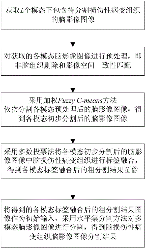
[0081] The present invention proposes a method for image segmentation of damaged lesion tissue in multimodal brain imaging, such as figure 2 shown, including the following steps:

[0082] Step 1: Obtain brain imaging images containing damaged lesion tissue to be segmented under L modalities.

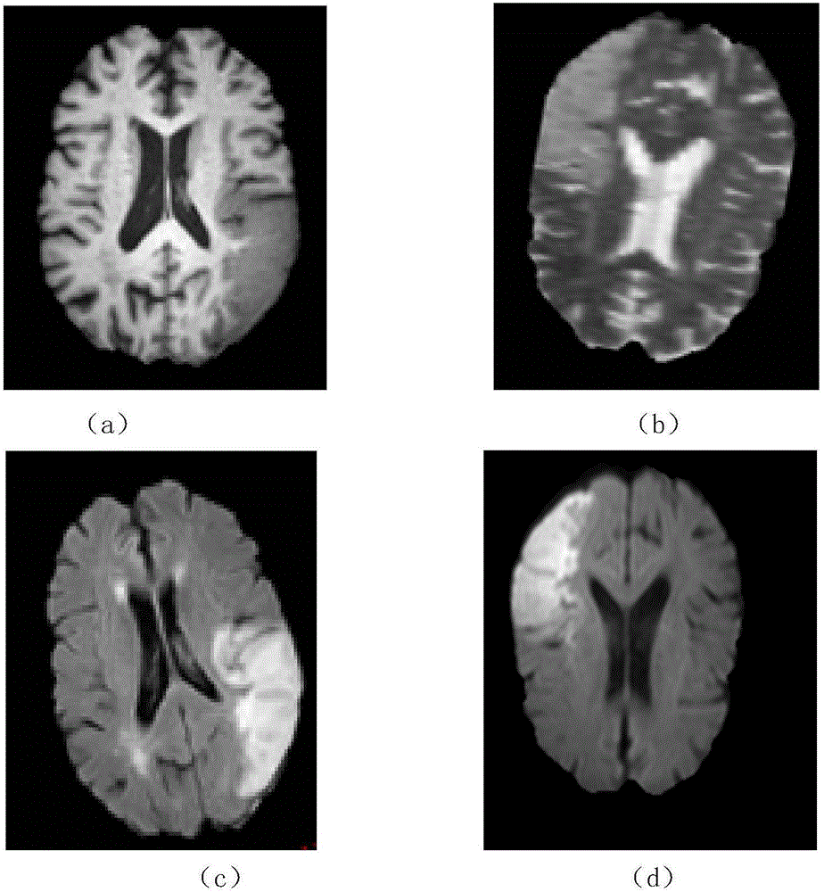
[0083] In this embodiment, brain imaging images containing damaged lesion tissue to be segmented under L=4 modalities are acquired, such as figure 1 shown.

[0084] Step 2: Perform preprocessing on the brain imaging images of each modality acquired in step 1, that is, non-brain tissue removal and image spatial consistency matching.

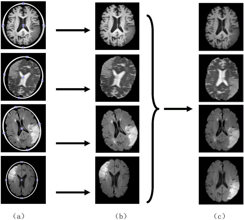
[0085] In this embodiment, there are two processing methods for preprocessing the brain imaging images of each modality acquired in step 1. The methods are as follows: image 3 shown, including the following steps: ...


PUM


Abstract

The invention provides a multi-modal brain-image injured pathological tissue image segmentation method, and the method comprises the steps: obtaining brain images comprising to-be-segmented injured pathological tissues at L modals; carrying out the preprocessing of the brain image at each modal; sequentially segmenting the brain images at all modals through employing a weighted Fuzzy C-means method after preprocessing; carrying out the label fusion of injured pathological tissues in the brain images at all modals after preliminary segmentation through employing a majority voting method, and obtaining a roughly segmented result image at each modal after label fusion; enabling the obtained roughly segmented result images at all modals as the initial input after label fusion, segmenting the multi-modal brain image through employing a horizontal set segmenting method, and obtaining a segmentation result of the injured pathological tissue image. The method can achieve the effective fusion of the beneficial information of a plurality of image modals, and avoids the negative effects on the image segmenting precision from a weak edge, image noise and inconsistent gray.

Description

technical field [0001] The invention belongs to the technical fields of computer vision, pattern recognition, image processing and analysis, and in particular relates to a method for segmenting images of damaged lesion tissues in multimodal brain images. Background technique [0002] The human brain is an important human organ that distinguishes human beings from other creatures and shows advanced intelligence after a long natural evolution. The structure of the human brain is complex, and corresponding lesions (such as cerebral apoplexy) have the characteristics of high incidence, high recurrence rate, high disability rate, and high fatality rate, which has become an important disease that endangers human health. Statistics show that stroke has become the second leading cause of death in the world. In my country, brain lesions seriously affect people's health and quality of life. According to the "Global Burden of Disease Report 2013" recently published by The Lancet, str...

Claims


Application Information

Patent Timeline
Patent Type & Authority Applications(China)
IPC IPC(8): G06T7/00G06T7/11G06T7/194G06K9/62
CPCG06T7/0012G06T2207/30016G06T2207/10088G06T2207/30096G06F18/2321
Inventor 冯朝路胡扬
Owner NORTHEASTERN UNIV
Why Eureka
Try Eureka