Portable non-mydriatic fundus imaging device

An imaging device and non-mydriatic technology, which is applied in the fields of eye testing equipment, ophthalmoscope, medical science, etc., can solve the problems of not providing infrared preview function, patient discomfort, and unsatisfactory shooting range, etc.

Inactive Publication Date: 2017-05-10
SHANGHAI MEDIWORKS PRECISION INSTR CO LTD
View PDF14 Cites 8 Cited by
  • Summary
  • Abstract
  • Description
  • Claims
  • Application Information

AI Technical Summary

Problems solved by technology

However, it does not provide an infrared preview function, but uses white light to directly irradiate the patient's fundus for a long

Method used


Image

  • Portable non-mydriatic fundus imaging device
  • Portable non-mydriatic fundus imaging device
  • Portable non-mydriatic fundus imaging device

Examples


Embodiment Construction

[0035] The present invention will be described in detail below in conjunction with specific embodiments. The following examples will help those skilled in the art to further understand the present invention, but do not limit the present invention in any form. It should be noted that those skilled in the art can make several changes and improvements without departing from the concept of the present invention. These all belong to the protection scope of the present invention.

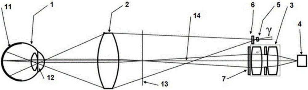
[0036] The present invention includes an infrared lighting source for black and white preview of the fundus, a white light flash light source for photographing fundus color photos, an eye-connecting objective lens assembly shared by the lighting system and the imaging system, and the second-order real image formed by the eye-connecting objective lens and the human eye optical system. A relay lens assembly for secondary imaging, and a two-dimensional image sensor that converts the secondary image into an ...


PUM

PropertyMeasurementUnit
Angleaaaaaaaaaa

Abstract

The invention provides a portable non-mydriatic fundus imaging device. The device comprises an eyepiece and a relay lens assembly which are sequentially arranged in front of a to-be-tested eye, and the eyepiece is located between the to-be-tested eye and the relay lens assembly. An imaging polarizer is arranged on the imaging light-in side of the relay lens assembly and between the eyepiece and the relay lens assembly. An image sensor assembly is arranged on the imaging light-out side of the relay lens assembly. The eyepiece, the imaging polarizer, the relay lens assembly and the image sensor assembly are coaxially arranged. An illumination assembly is transversely arranged on one side of an optical axis in a deviated mode. The device has the advantages that under the condition that the to-be-tested eye is subjected to mydriasis without medicine, the to-be-tested eye can be observed in real time and subjected to video recording and flash photography. The device is mainly characterized in that high-definition imaging of the fundus of the to-be-tested eye is achieved by means of stray light, the host image subduction technology and the aberration correction technology on the premise that portability is maintained.

Description

technical field [0001] The invention relates to an imaging device, which belongs to the technical field of medical instrument microscopes, in particular to a portable non-mydriatic fundus imaging device. Background technique [0002] The fundus camera is a traditional medical device, which is used to observe and record the retinal images of the human eye, and provide objective basis for doctors to analyze and diagnose. The only capillaries that can be directly observed in the human body are distributed on the retina detected by the fundus camera. The changes in the shape of these capillaries can be used for early diagnosis of fundus diseases, hypertension, diabetes and other diseases, and have become an important means of modern medical diagnosis. . The world's first desktop fundus camera was successfully trial-produced by the German CARLZEISS company in 1925. Subsequently, CANON, TOPCON, NIKON, OLYMPUS, OPTON and other companies made a lot of improvements to the design of ...

Claims


Application Information

Patent Timeline
IPC IPC(8): A61B3/12A61B3/14
CPCA61B3/12A61B3/0008A61B3/0016A61B3/14
Inventor 杨丛渊陈文光郭丁华赵立秀魏悦赵小飞皮晓奔
Owner SHANGHAI MEDIWORKS PRECISION INSTR CO LTD
Why Eureka
Try Eureka