A colorimetric immunoassay for the detection of tumor markers for non-diagnostic purposes

A tumor marker and immunoassay technology, applied in the field of colorimetric immunoassay, can solve the problems of short reaction time, expensive equipment, complicated operation, etc., and achieve the effect of simple operation, fast response and low cost

Active Publication Date: 2018-06-22
上海塞力斯医学检验实验室有限公司
View PDF6 Cites 0 Cited by
  • Summary
  • Abstract
  • Description
  • Claims
  • Application Information

AI Technical Summary

Problems solved by technology

[0005] The purpose of the present invention is to provide a colorimetric immunoassay method for detecting tumor markers for non-diagnostic purposes in view of the problems of expensive instruments for detecting tumor markers, complicated operations, and high cost of detection reagents. The method is easy to operate and low in cost. Inexpensive, does not require expensive instruments and reagents, high sensitivity, short reaction time

Method used


Image

  • A colorimetric immunoassay for the detection of tumor markers for non-diagnostic purposes
  • A colorimetric immunoassay for the detection of tumor markers for non-diagnostic purposes
  • A colorimetric immunoassay for the detection of tumor markers for non-diagnostic purposes

Examples


Embodiment 1

[0021] Example 1 A colorimetric immunoassay method for detecting the tumor marker CEA for non-diagnostic purposes, comprising the following steps:

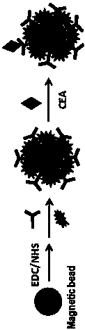
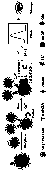
[0022] (1) Preparation of functionalized magnetic beads

[0023] Take 4μL of 25mg / mL carboxylated magnetic beads, under the action of an external magnetic field, wash three times with 50mM tris(hydroxymethyl)aminomethane-hydrochloric acid buffered saline solution, pH=7.4, and add to the washed magnetic beads Mix 200 μL of 0.2M 1-ethyl-(3-dimethylaminopropyl) carbodiimide hydrochloride (EDC) and 0.2M N-hydroxysuccinimide (NHS), shake at room temperature React for 30 minutes to activate the carboxylic acid groups on the surface of the magnetic beads. After the activated magnetic beads were washed three times with 50mM Tris-HCl solution, they were redispersed in 400μL of 2-(N-morpholine)B with a concentration of 50mM and pH=6.0. Add 10 μL of carcinoembryonic antigen CEA-coated antibody at a concentration of 1 mg / mL to the sulfonic a...

Embodiment 2

[0026] The detection of CEA content in the embodiment 2 standard solution

[0027]Take 40 μL of the prepared functionalized magnetic beads and place them in 6 sample tubes, dilute the CEA standard solution with a concentration of 1 mg / mL with 50 mM Tris-HCl solution at pH=7.4 to a concentration gradient of 0.001, 0.01, 0.1, 1, 10, and 100ng / mL CEA standard stock solution, put 100 μL of the above six different concentrations of CEA standard stock solution in the above six sample tubes in turn, and incubate at 37°C for 20 minutes with shaking. Under the action of an external magnetic field, use Tris-HCl solution containing 0.05% (w / v) Tween at a concentration of 50 mM, pH = 7.4 and Tris-HCl solution were washed alternately, and then 100 μL of urease functionalized gold nanoprobe was added to each sample tube respectively, Shake and incubate at 37°C for 20 minutes, wash the product three times under the action of an external magnetic field, and then add urea with a concentration ...

Embodiment 3

[0030] Example 3 Detection of CEA content in human serum

[0031] Get three clinical serum samples, carry out 5 parallel experiments respectively for each sample, the processing method of sample is the same as above-mentioned standard solution, see the following table 2 through the concentration of CEA in the tested sample, use the present embodiment method of above-mentioned three clinical serum samples Compared with the measurement results of the Beckman chemiluminescence immunoassay analyzer, the results are shown in Table 2 below.

[0032] Table 2 Comparison of the measurement results between this example and the Beckman Chemiluminescent Immunoassay Analyzer

[0033]

[0034] As can be seen from Table 2 above, the relative standard deviation (RSD) of the test results in this example is 1.8-5.6%, and the relative error is -3.7-3.1%, indicating that the immunoassay method of this example has high repeatability and stability. Compared the test results of this embodiment ...


PUM


Abstract

The invention discloses a colorimetric immunoassay method convenient for detecting a tumor marker. The colorimetric immunoassay method comprises preparation of a functional magnetic bead, preparation of a urease functional gold-nano probe and detection of contents of CEA in a standard solution and a sample. The method can be also used for detecting alpha fetal proteins (AFP), pancreatic oncofetal antigens (POA), isoenzyme (NSE), hormone (HCG) and the like. The method has the characteristics of simple operation, low cost and quick reaction; quantitative detection of an object to be detected can be carried out with only eyes or a simple ultraviolet-visible spectrophotometer, so that the colorimetric immunoassay method has an important significance for popularization of actual application of colorimetric immunoassay to aspects such as clinical diagnosis, medicine analysis and food safety.

Description

technical field [0001] The invention relates to the technical field of colorimetric immunoassay, in particular to a colorimetric immunoassay method for detecting tumor markers for non-diagnostic purposes. Background technique [0002] Immunoassay is a highly selective and sensitive analysis method developed based on the highly specific immune recognition between antigen and antibody. The theoretical basis of immune analysis is the specific reaction between antigen and antibody, that is, immune response. Colorimetric immunoassays generally use antibodies or antigens as biological sensitive elements. After immobilizing them on the surface of some carriers, the tracer markers can be captured quantitatively through the antigen-antibody immune reaction on the carrier surface, and then the tracer markers can be combined with the Based on the color reaction of specific detection reagents to generate colored compounds, a quantitative analysis relationship is established by comparin...

Claims


Application Information

Patent Timeline
Patent Type & Authority Patents(China)
IPC IPC(8): G01N33/574
CPCG01N33/57484
Inventor 赖国松黎波胡胜利
Owner 上海塞力斯医学检验实验室有限公司
Why Eureka
Try Eureka