Sub-pixel-based blood vessel segmentation method and segmentation system

A sub-pixel and sub-pixel edge technology is applied in the field of sub-pixel-based blood vessel segmentation methods and segmentation systems. , the edge of blood vessels is clear, the effect of improving the display effect

Inactive Publication Date: 2017-09-12
CENT SOUTH UNIV
View PDF3 Cites 10 Cited by
  • Summary
  • Abstract
  • Description
  • Claims
  • Application Information

AI Technical Summary

Problems solved by technology

In view of the complex morphological structure of blood vessels, as well as the noise in the imaging environment, the attenuation and uneven distribution of contrast agents, etc., there are often various problems in ...

Method used


Image

  • Sub-pixel-based blood vessel segmentation method and segmentation system
  • Sub-pixel-based blood vessel segmentation method and segmentation system
  • Sub-pixel-based blood vessel segmentation method and segmentation system

Examples


Embodiment Construction

[0045]The technical solutions in the embodiments of the present invention will be clearly and completely described below. Obviously, the described embodiments are only some of the embodiments of the present invention, not all of them. Based on the embodiments of the present invention, all other embodiments obtained by persons of ordinary skill in the art without making creative efforts belong to the protection scope of the present invention.

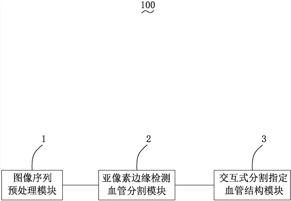
[0046] see figure 1 , the present invention provides a sub-pixel-based blood vessel segmentation system 100, including an image sequence preprocessing module 1, a sub-pixel edge detection blood vessel segmentation module 2, and an interactive segmentation and specified blood vessel structure module 3.

[0047] The image sequence preprocessing module 1 is used to preprocess the Dicom file sequence obtained by CT scanning through high-order interception, inverse transformation and window adjustment, and convert the Dicom file sequence into...


PUM


Abstract

The invention provides a sub-pixel-based blood vessel segmentation method. The method comprises: S1, a Dicom file sequence is converted to a Bmp file sequence that is universal for a computer; S2, a rough edge detection result is generated; S3, the rough edge detection result is generated into a linked list and the linked list is recorded; S4, an edge coordinate is obtained; S5, a three-dimensional blood vessel structure is obtained; and S6, regional growth segmentation is carried out on the three-dimensional blood vessel structure by a 26-neighbourhood way to obtained a designated blood vessel structure by interactive segmentation. In addition, the invention also provides a sub-pixel-based blood vessel segmentation system. Compared with the prior art, the sub-pixel-based blood vessel segmentation method and system have the following beneficial effects: a border crossing phenomenon in Dicom file window modulation processing can be avoided and the display effect can be improved; a problem of difficult threshold selection in rough edge detection can be solved; the blood vessel edge becomes clear and the details are perfected; blood vessel segmentation can reach sub-pixel-level precision; and interactive segmentation of the designated blood vessel structure can be realized.

Description

technical field [0001] The present invention relates to the technical field of blood vessel segmentation, in particular to a subpixel-based blood vessel segmentation method and a segmentation system thereof. Background technique [0002] In recent years, with the rapid development of biomedical imaging technology and medical imaging equipment, the resolution of medical images has been increasing day by day, making medical imaging one of the fastest-growing fields in medical technology. Therefore, the role and influence of medical image processing technology in medical research and clinical practice is extremely important. Medical image segmentation is an important application of image segmentation technology in the medical field and an important part of medical image processing, which can provide doctors with important clinical reference value. In medical image segmentation, blood vessel segmentation has always been a research hotspot. [0003] With the improvement of huma...

Claims


Application Information

Patent Timeline
IPC IPC(8): G06T7/00G06T7/11G06T7/13G06T7/136
CPCG06T7/0012G06T2207/10081G06T2207/30101
Inventor 廖胜辉任巧丽李建锋腾光禹任辉贺佳丽邹北骥
Owner CENT SOUTH UNIV
Why Eureka
Try Eureka