
Partitioning algorithm for choroidal neovascularization in OCT image

A new blood vessel, segmentation algorithm technology, applied in image analysis, image enhancement, image data processing and other directions, can solve the problems of affecting segmentation accuracy, not taking into account, limiting model segmentation performance, etc., to enhance correlation and improve segmentation accuracy. Effect

Active Publication Date: 2017-11-21
SUZHOU BIGVISION MEDICAL TECH CO LTD
View PDF7 Cites 23 Cited by
  • Summary
  • Abstract
  • Description
  • Claims
  • Application Information

AI Technical Summary

Problems solved by technology

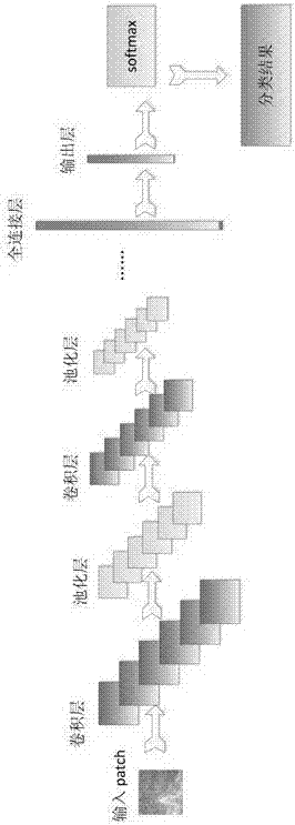
However, due to the complex characteristics of OCT images, there are two shortcomings in the direct use of convolutional neural networks to segment choroidal neovascularization: (1) The traditional method first needs to divide the image into several small patches (patch), and then train the convolution based on the patch. Neural Network Segmentation Model
However, there are some structural correlations (such as local similarity) between patches and patches. Traditional methods do not take this effective structural information into account when training convolutional neural network segmentation models, thus limiting the segmentation performance of the model.
(2) The traditional model is based on a single scale patch
The size of choroidal neovascularization is not fixed, so it is difficult for a single-scale patch to obtain effective context information, which affects the segmentation accuracy

Method used


Image

  • Partitioning algorithm for choroidal neovascularization in OCT image
  • Partitioning algorithm for choroidal neovascularization in OCT image
  • Partitioning algorithm for choroidal neovascularization in OCT image

Examples


Embodiment 1

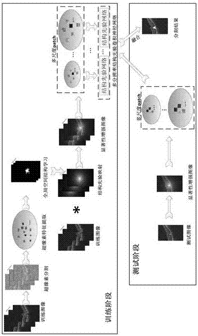
[0041] A choroidal neovascularization segmentation algorithm in OCT images, such as figure 1 As shown, this method mainly includes two stages: training stage and testing stage, the specific steps are as follows:

[0042] (1) The training phase mainly includes two parts: "structural prior learning" and "multi-scale structural prior convolutional neural network training". The specific steps are as follows:

[0043] S01: Design a structure prior learning method for the training image, construct a structure prior matrix, and the structure prior matrix is ​​used to distinguish the choroidal neovascularization area and the background area;

[0044] Described structure prior learning method comprises the following steps:

[0045] a: Perform superpixel segmentation on the training image to obtain several superpixel regions, and use the SLIC algorithm to segment;

[0046] b: extract features, the features include the average gray value of each superpixel, texture features based on co...


PUM


Abstract

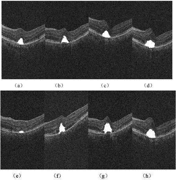
The invention discloses a partitioning algorithm for choroidal neovascularization in an OCT image. The algorithm includes steps of S01, designing a structure prior learning method for a training image and constructing a structure prior matrix used for distinguishing a choroidal neovascularization zone and a background zone; S02, converting the OCT original image to a saliency enhancing image used for enhancing the saliency of the choroidal neovascularization zone based on the structure prior matrix; S03, adopting multi-scale analysis on the saliency enhancing image and dividing the saliency enhancing image into m scales; S04, acquiring m trained convolutionneural network model based on each scale training; S05, processing a testing image by utilizing the step S01, S02, S03 and performing testing by utilizing the trained convolutionneural network model in the step S04, outputting m portioning results and fusing the m partitioning results into the final partitioning result. By adopting the algorithm provided by the invention, the precision of partitioning of the choroidal neovascularization in the OCT image can be improved distinctively.

Description

technical field [0001] The invention relates to a choroidal neovascularization segmentation algorithm in an OCT image, belonging to the technical field of retinal image segmentation. Background technique [0002] Most of the existing automatic segmentation techniques for choroidal neovascularization are based on fundus fluorescein angiography images. Compared with fundus fluorescein angiography, OCT images have the advantages of non-invasive, high-speed, high-resolution, three-dimensional imaging, etc., and it has more important clinical significance for the auxiliary diagnosis of common clinical ophthalmic diseases such as age-related macular degeneration. [0003] Currently, there is no choroidal neovascularization segmentation algorithm based on OCT images. The segmentation of choroidal neovascularization in OCT images faces many challenges: large texture changes, gray heterogeneity, inconsistent shape and size, blurred boundaries, and a large amount of speckle noise. T...

Claims


Application Information

Patent Timeline
Patent Type & Authority Applications(China)
IPC IPC(8): G06T7/11G06T7/194G06K9/46G06K9/62
CPCG06T7/11G06T7/194G06T2207/20081G06T2207/30101G06V10/462G06F18/23213
Inventor 陈新建袭肖明
Owner SUZHOU BIGVISION MEDICAL TECH CO LTD
Learn More