A percutaneous nephroscopic dilation sheath

A nephroscopic and dilating tube technology, applied in dilators, medical science, surgery, etc., can solve the problems of inability to observe the dilatation sheath, renal hemorrhage, and deep puncture, and achieve the effect of preventing renal hemorrhage.

Active Publication Date: 2022-03-29
蓝线铂立生命科技(苏州)有限公司
View PDF9 Cites 0 Cited by
  • Summary
  • Abstract
  • Description
  • Claims
  • Application Information

AI Technical Summary

Problems solved by technology

[0003] However, the traditional percutaneous nephroscope still has defects. For example, when the expansion is performed after the puncture, the traditional expansion sheath cannot observe the position of the expansion sheath, which may easily cause medical accidents such as excessive puncture and renal hemorrhage.

Method used


Image

  • A percutaneous nephroscopic dilation sheath
  • A percutaneous nephroscopic dilation sheath
  • A percutaneous nephroscopic dilation sheath

Examples


Embodiment Construction

[0019] In order to make the technical means, creative features, goals and effects achieved by the present invention easy to understand, the present invention will be further described below in conjunction with specific illustrations.

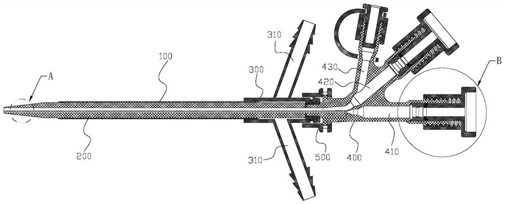
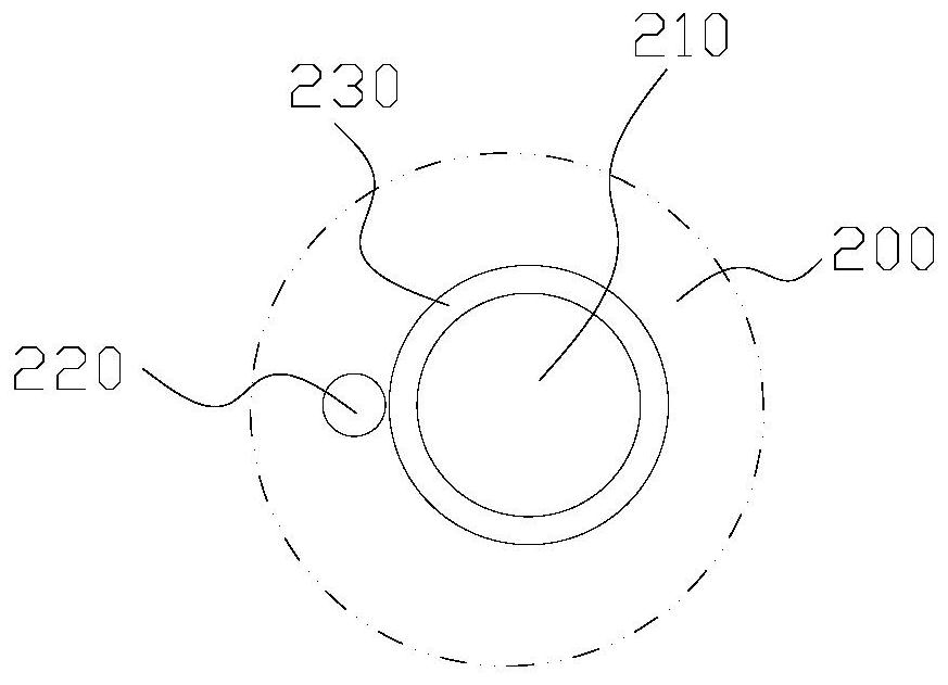
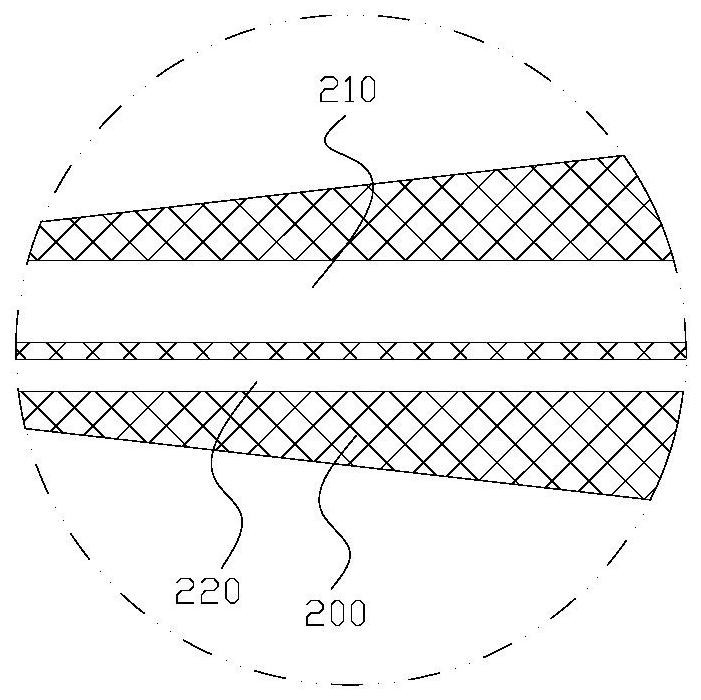
[0020] see Figure 1-Figure 3 , a dilation sheath for percutaneous nephroscopic visualization, comprising a tearing sheath 100 and a dilation tube 200 arranged in the tearing sheath 100, the dilation tube 200 is cylindrical, the front end is a cone, and the top of the cone is a circular plane 230 , the dilation tube 200 is provided with a fiber channel 210 and a guide wire channel 220 . Wherein the optical fiber channel 210 can pass through the optical fiber and inject water, and the guide wire channel 220 can pass through the guide wire. The fiber channel 210 and guide wire channel 220 are arranged parallel to the expansion tube 200 , and the fiber channel 210 passes through the circular plane 230 , the guide wire channel 220 passes through th...


PUM


Abstract

The invention relates to a percutaneous nephroscopic expansion sheath, which comprises a tear sheath and an expansion tube arranged in the tear sheath. The expansion tube is cylindrical, with a conical front end, and the top of the cone is a circular plane. A fiber optic channel and a guide wire channel are provided, and the fiber channel and the guide wire channel are arranged parallel to the expansion tube, and the fiber channel passes through the circular plane, and the guide wire channel passes through the outer surface of the cone, and the fiber channel is concentric with the circular plane. The beneficial effect of the present invention is that the visual image system can be used to effectively locate the puncture expansion when the expansion puncture is performed by using the visible expansion sheath, so as to prevent medical accidents such as renal hemorrhage caused by too deep puncture in conventional operations.

Description

technical field [0001] The invention relates to a medical device, in particular to a percutaneous nephroscope. Background technique [0002] Surgical treatment methods for kidney stones include: open lithotomy, extracorporeal lithotripsy, and percutaneous nephrolithotomy. The traditional method of "removing stones with a knife" requires a large incision of about 20 cm in the waist, incision of the muscles in the waist, separation of the kidneys, and a small incision in the renal pelvis or renal parenchyma to remove the stones. This method causes more damage, more bleeding, more pain, and slower recovery. Usually you need to stay in the hospital for about 7 days after the operation. Because the surgical incision is large, not only the skin is not beautiful, but also the physical strength of the patient is greatly affected after the operation. Compared with open surgery, percutaneous nephrolithotomy has the advantages of less damage, less pain, complete stone removal, and f...

Claims


Application Information

Patent Timeline
Patent Type & Authority Patents(China)
IPC IPC(8): A61B17/22A61B17/00A61B17/34A61M29/00
CPCA61M29/00A61B17/00234A61B17/22A61B17/3403A61B17/3494A61B2017/22038A61B2017/22079A61B2017/22082A61B2017/3405A61M2210/1082A61M2210/005
Inventor 程跃刘冠琳史军谢国海严泽军郁鹏忠
Owner 蓝线铂立生命科技(苏州)有限公司
Why Eureka
Try Eureka