Image attenuation correction method and device

An attenuation correction and image technology, applied in computerized tomography scanners, echo tomography, etc., can solve problems such as increasing the misdiagnosis rate of doctors, affecting the matching between PET images and CT images, and attenuation artifacts in PET images, and achieving improved The effect of image quality

Active Publication Date: 2018-01-30
JIANGSU SINOGRAM MEDICAL TECH CO LTD
View PDF6 Cites 23 Cited by
  • Summary
  • Abstract
  • Description
  • Claims
  • Application Information

AI Technical Summary

Problems solved by technology

However, various physiological processes such as the subject's breathing and/or heartbeat can affect the matching of PET and CT images even without taking into account the voluntary or controlled movements that the subject may be making
When the matching between the PET image and the CT image

Method used


Image

  • Image attenuation correction method and device
  • Image attenuation correction method and device
  • Image attenuation correction method and device

Examples


Embodiment Construction

[0025] In order to make the objectives, technical solutions, and advantages of the present invention clearer, the embodiments of the present invention will be described in further detail below in conjunction with the accompanying drawings.

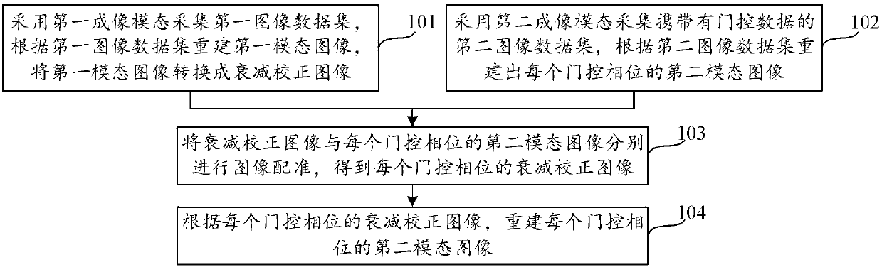
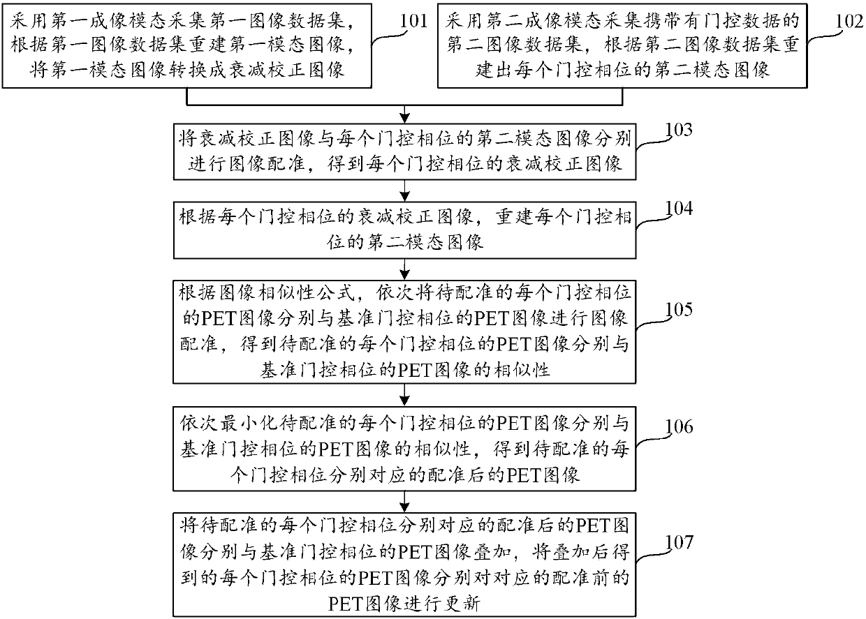
[0026] Figure 1A Is a flowchart of an image attenuation correction method provided in an embodiment of the present invention, such as Figure 1A As shown, the image attenuation correction method includes the following steps.

[0027] Step 101: Collect a first image data set using a first imaging modality, reconstruct a first modality image according to the first image data set, and convert the first modality image into an attenuation correction image.

[0028] It should be noted that the first imaging modality is one of Computed Tomography (CT) or Magnetic Resonance Imaging (MRI).

[0029] Step 102: Use the second imaging modality to collect a second image data set carrying gated data, and reconstruct a second modality image of each gated phase acco...


PUM


Abstract

The invention discloses an image attenuation correction method and device and belongs to the technical field of medical imaging. The image attenuation correction method comprises the steps that a first mode image is reconstructed according to first image data and is converted into an attenuation correction image; a second mode image of each gating phase is reconstructed according to second image data; image registration is conducted on the attenuation correction image and the second mode image corresponding to each gating phase to obtain an attenuation correction image of each gating phase; according to the attenuation correction image of each gating phase, the second mode image of each gating phase is reconstructed. The problem is solved that in the relevant art, the matching of PET and CT images is reduced due to the movement in the examination process or the physiological process of a subject, accordingly attenuation correction malposition of the PET images is likely to produce andattenuation artifacts are produced in the PET images, and the advantages of attenuation correction of PET data and quality improvement of PET images are achieved.

Description

Technical field [0001] The embodiments of the present invention relate to the technical field of medical image imaging, and in particular to an image attenuation correction method and device. Background technique [0002] Positron Emission Tomography (Positron Emission Tomography, PET) is a three-dimensional imaging non-destructive detection technology that uses positron radioisotope-labeled compounds to be injected into the body, and their spatial distribution and time characteristics are measured in vitro, with high sensitivity , Good accuracy and accurate positioning. [0003] The working principle of PET is: labeling a positron-emitting radionuclide to a compound that can participate in the blood flow or metabolic process of human tissue, and injecting the radionuclide labeled with a positron-bearing compound into the subject, allowing the subject to be tested The person performs PET imaging within the effective field of view of PET. During the PET scanning process, the posit...

Claims


Application Information

Patent Timeline
IPC IPC(8): A61B6/03
Inventor 朱程李楠
Owner JIANGSU SINOGRAM MEDICAL TECH CO LTD
Why Eureka
Try Eureka