Deep-network-based tissue segmentation method of panoramic digital colorectum pathology image

A digital pathology, deep network technology, applied in the field of medical image processing, can solve the problems of inaccurate processing results, small scope of application, poor accuracy, etc.

Active Publication Date: 2018-02-06
NANJING UNIV OF INFORMATION SCI & TECH
View PDF5 Cites 21 Cited by
  • Summary
  • Abstract
  • Description
  • Claims
  • Application Information

AI Technical Summary

Problems solved by technology

Histopathological images are highly complex and have many targets. Existing research work on colorectal digital pathology images is very little. Generally, only some types of tissue areas in the images are detected, and the images are processed one-sidedly, and the processing results are not accurate. precise
[0004] Existing research on colorectal panoramic digital pathological images has not yet done research on colorectal panoramic digit...

Method used


Image

  • Deep-network-based tissue segmentation method of panoramic digital colorectum pathology image
  • Deep-network-based tissue segmentation method of panoramic digital colorectum pathology image
  • Deep-network-based tissue segmentation method of panoramic digital colorectum pathology image

Examples


Embodiment Construction

[0035] The present invention will be further described below in conjunction with the accompanying drawings.

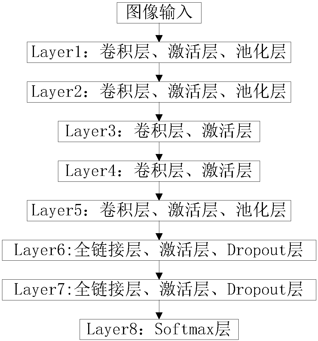
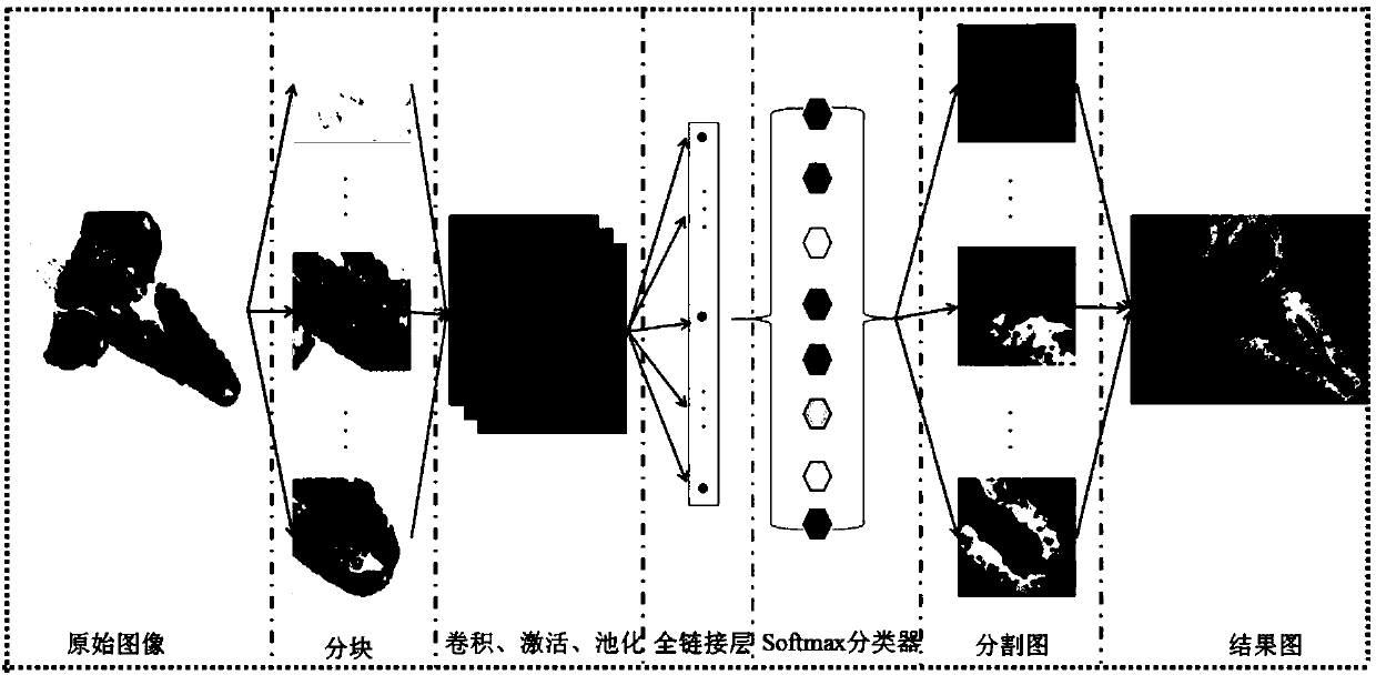
[0036] Such as figure 1 and figure 2 As shown, the tissue segmentation method of colorectal panoramic digital pathology image based on deep network includes the following steps:

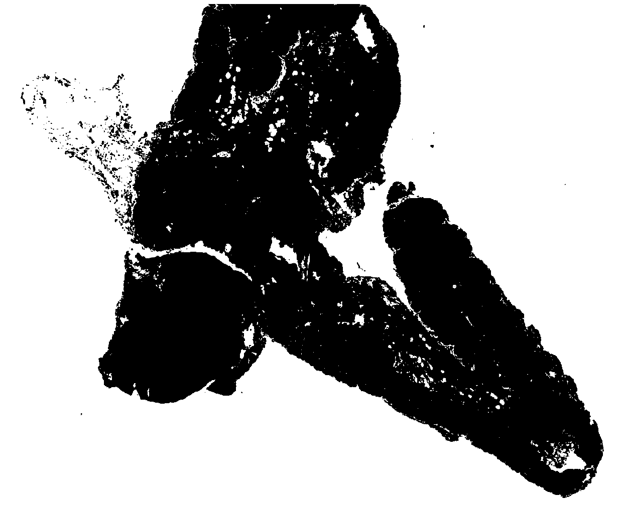
[0037] (1) Acquire colorectal panoramic digital pathology pictures under a magnifying glass: select the panoramic digital colorectal pathology data under a magnifying glass of 20 times; image 3 for the original image, Figure 4 It is divided into 5000*5000 size images under 20 times.

[0038] (2) Segment the panoramic digital image of the colorectum into 5000*5000 segmented images, all segmented images retain the block coordinates in the panoramic digital image, and use the sliding window and the trained model to mark the tissue types in turn for all segmented images, Get each 5000×5000 segmented image marked with tissue type;

[0039] Step (2) specifically comprises the following step...


PUM


Abstract

The invention discloses a deep-network-based tissue segmentation method of a panoramic digital colorectum pathology image. The method comprises the following steps: step one, acquiring a panoramic digital colorectum pathology image; step two, segmenting the panoramic digital colorectum pathology image; step three, establishing a training sample image; step four, extracting deep features of tissuesof different types; step five, determining types of tissues in the segmented images by using a classifier based on the extracted deep tissue features; step six, splicing image classification resultsand determining a tissue class of an overall picture; and step seven, splicing the images according to block coordinates. According to the invention, the panoramic digital colorectum pathology image is segmented and tissue type marking is carried out on all segmented images by using a sliding window and a trained model; and tissue type determination is carried out by using the classifier based onthe extracted deep tissue features to obtain an image classification result. The classification becomes accurate and the classification speed is increased.

Description

technical field [0001] The invention discloses a tissue segmentation method for colorectal panorama digital pathological images based on a deep network, belonging to the field of medical image processing. Background technique [0002] At present, the analysis of pathological images is mainly evaluated by pathologists. However, the manual analysis method is extremely time-consuming and involves the subjective judgment of doctors. There are large differences between doctors with different experiences, which will lead to inappropriate treatment or overtreatment. In poorer and backward areas, many people died due to missing treatment time due to lack of good doctors and medical equipment. [0003] For pathological tissue images, since they contain a lot of valuable information, different histopathological images can be classified by using some characteristics of the pathological image itself. Histopathological images are highly complex and have many targets. Existing research ...

Claims


Application Information

Patent Timeline
IPC IPC(8): G06T7/11G06T3/40G06K9/62G06N3/08G06N3/04
CPCG06N3/08G06T3/4038G06T7/11G06T2207/10056G06T2207/20084G06T2207/20081G06T2207/30028G06T2207/30096G06N3/048G06F18/24
Inventor 徐军蔡程飞徐海俊孙明建
Owner NANJING UNIV OF INFORMATION SCI & TECH
Why Eureka
Try Eureka