DNA (desoxyribonucleic acid) complex structure variation diagnosis method

A diagnostic method and technology of complex structure, applied in the field of gene detection of tumor cells, can solve the problems of complex structure mutation diagnosis and chromosomal structure mutation technology, such as slow speed and inability, to save time and cost, avoid obtaining wrong information, and improve the effect of accuracy

Active Publication Date: 2018-06-15
志诺维思(北京)基因科技有限公司
View PDF4 Cites 2 Cited by
  • Summary
  • Abstract
  • Description
  • Claims
  • Application Information

AI Technical Summary

Problems solved by technology

[0003] The purpose of the present invention is to provide a method for diagnosing complex structural variations of DNA to solve the problems that existing techniques for chromosomal structural variations are slow and unable to diagnose complex structural variations

Method used


Image

  • DNA (desoxyribonucleic acid) complex structure variation diagnosis method
  • DNA (desoxyribonucleic acid) complex structure variation diagnosis method
  • DNA (desoxyribonucleic acid) complex structure variation diagnosis method

Examples


Embodiment 1

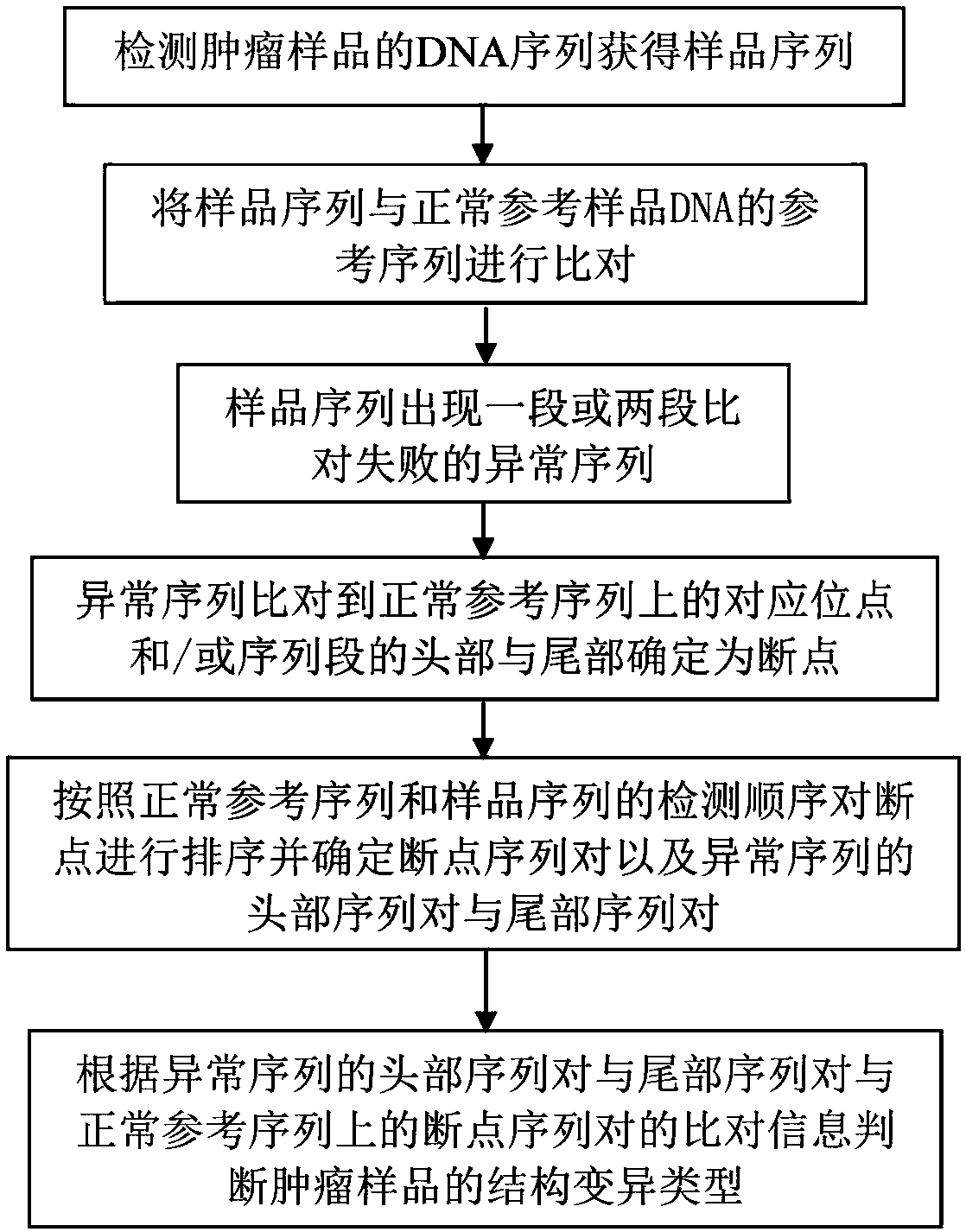
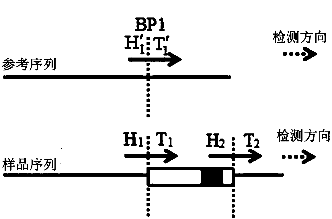
[0037] Such as figure 1 As shown, a DNA complex structure mutation diagnosis method disclosed in this embodiment includes: detecting the DNA sequence of the tumor sample to obtain the sample sequence; comparing the sample sequence with the reference sequence of the normal reference sample DNA; image 3 As shown, when the sample sequence is compared with the reference sequence of the normal reference sample DNA, there is an abnormal sequence in the sample sequence that fails to be aligned; the corresponding position of the abnormal sequence compared to the normal reference sequence is determined as the breakpoint BP1; according to the normal reference sequence Sort the breakpoints with the detection order of the sample sequence and determine the breakpoint sequence pair (H′ 1 , T 1 ′) and the head sequence pair (H 1 , T 1 ) And the tail sequence pair (H 2 , T 2 ); the head point H of the head sequence pair of the abnormal sequence on the sample sequence 1 The tail point T of the ta...

Embodiment 2

[0039] Such as figure 1 As shown, a DNA complex structure mutation diagnosis method disclosed in this embodiment includes: detecting the DNA sequence of the tumor sample to obtain the sample sequence; comparing the sample sequence with the reference sequence of the normal reference sample DNA; Figure 4 As shown, when the sample sequence is compared with the reference sequence of the normal reference sample DNA, there is a missing sequence in the sample sequence; this missing sequence is compared to the head and tail of the sequence segment to determine the breakpoint; according to the normal reference sequence and the sample sequence Sort the breakpoints BP1 and BP2 and determine the breakpoint sequence pair (H′ 1 , T 1 ′) and (H′ 2 , T 2 ′) and the sequence pair (H 1 , T 1 ); the head point H of the corresponding position of the missing sequence on the sample sequence 1 With tail point T 1 Align respectively to the head point H′ of the head breakpoint sequence pair on the normal...

Embodiment 3

[0041] Such as figure 1 As shown, a DNA complex structure mutation diagnosis method disclosed in this embodiment includes: detecting the DNA sequence of the tumor sample to obtain the sample sequence; comparing the sample sequence with the reference sequence of the normal reference sample DNA; Figure 5 As shown, when the sample sequence is compared with the reference sequence of the normal reference sample DNA, there is an abnormal sequence that fails the alignment; the head and tail of the sequence segment where the abnormal sequence is compared to the normal reference sequence are determined as breakpoints; Sequence the breakpoints BP1 and BP2 according to the detection sequence of the normal reference sequence and the sample sequence, and determine the breakpoint sequence pair (H′ 1 , T 1 ′) and (H′ 2 , T 2 ′) and the head sequence pair (H 1 , T 1 ) And the tail sequence pair (H 2 , T 2 ); the head point H of the head sequence pair of the abnormal sequence on the sample sequen...


PUM


Abstract

The invention discloses a DNA (desoxyribonucleic acid) complex structure variation diagnosis method. 16 chromosome structure variation types are designed, and a corresponding judgment model is designed by aiming at each variation type. Comparison information is directly obtained in a sequencing data comparison file. Compared with the prior art, the method omits sequence re-splicing and re-comparing parts, DNA complex structure variation detection can be quickly finished, and time cost is saved. Meanwhile, since information required for deducing the variation type is fully obtained in the comparison, the problem that re-comparison information can not be completely obtained and wrong information is obtained in a traditional process can be avoided, and chromosome structure variation detectionaccuracy is improved.

Description

Technical field [0001] The invention relates to the technical field of gene detection of tumor cells, in particular to a method for diagnosing complex DNA structural mutations. Background technique [0002] Existing chromosome structure variation detection technology uses traditional simple structure variation types. When looking for breakpoints, it lacks necessary information collection and judgment work, and does not classify complex variation types, such as chromosome structure variation detection software such as DELLY. There are only five types of structural variation. When judging the type of structural variation, it takes a lot of time and computing resources to retrieve the relevant information about the breakpoint. This traditional process of judging structural variations not only causes high time and resource costs, but also affects the accuracy of the diagnosis of structural variations. Therefore, the prior art cannot perform complex structural mutation diagnosis. S...

Claims


Application Information

Patent Timeline
Patent Type & Authority Applications(China)
IPC IPC(8): G06F19/20G06F19/18
CPCG16B20/00G16B25/00
Inventor 张忠波郝伶童尹龙辉曹丽华凌少平
Owner 志诺维思(北京)基因科技有限公司
Why Eureka
Try Eureka