Kit and method for diagnosis of lung cancer in chronic obstructive pulmonary disease (COPD) patients through meta-genome analysis

A kit, patient's technology, applied in the increase or field of content

Inactive Publication Date: 2018-07-03
MD HEALTHCARE INC
View PDF0 Cites 9 Cited by
  • Summary
  • Abstract
  • Description
  • Claims
  • Application Information

AI Technical Summary

Problems solved by technology

However, regarding the occurrence of lung cancer, there is no report on a method for identifying the causative agent of lung cancer from human fluids (such as blood, urine, etc.)

Method used


Image

  • Kit and method for diagnosis of lung cancer in chronic obstructive pulmonary disease (COPD) patients through meta-genome analysis
  • Kit and method for diagnosis of lung cancer in chronic obstructive pulmonary disease (COPD) patients through meta-genome analysis
  • Kit and method for diagnosis of lung cancer in chronic obstructive pulmonary disease (COPD) patients through meta-genome analysis

Examples


Embodiment 1

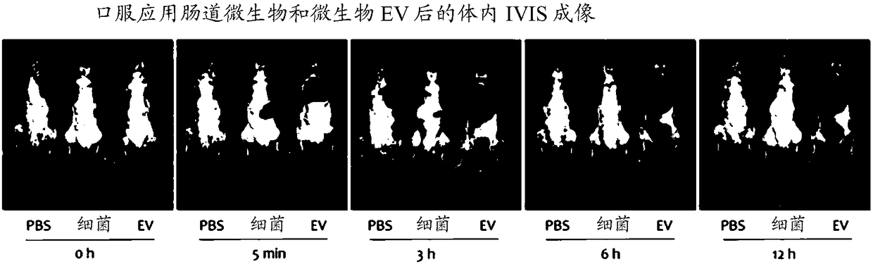
[0084] Example 1. Analysis of in vivo absorption, distribution and excretion patterns of enteric bacteria and bacteria-derived vesicles

[0085] In order to evaluate whether enteric bacteria and bacteria-derived extracellular vesicles are systematically absorbed through the gastrointestinal tract, experiments were performed using the following methods. More specifically, 50μg each of fluorescently labeled Pseudomonas enterobacteria and extracellular vesicles (EV) derived from the bacteria were orally administered to the gastrointestinal tract of mice, and at 0 hours and at Fluorescence was measured after 5 minutes, 3 hours, 6 hours and 12 hours.

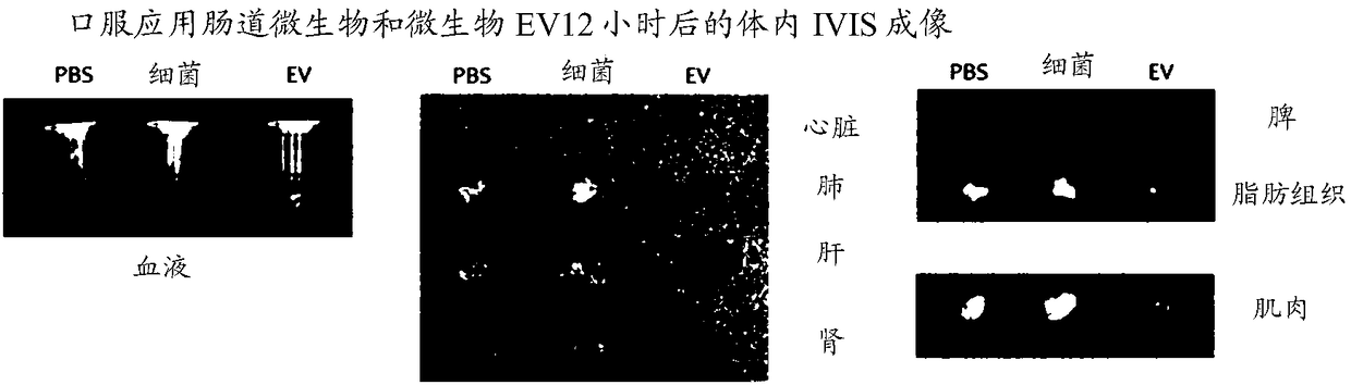
[0086] As a result of observing the overall image of the mouse, such as Figure 1A As shown, bacteria are not absorbed by the system during administration, and EVs derived from bacteria are distributed in the lungs 5 ​​minutes after administration, and strong fluorescence is observed in the bladder 3 hours after administration, confirming ...

Embodiment 2

[0088] Example 2. Vesicle isolation and DNA extraction from blood

[0089] In order to separate vesicles from blood and extract DNA, first add serum to a 10ml tube, centrifuge at 3,500×g and 4°C for 10 minutes to precipitate the suspension, and collect only the supernatant, then place it in In a new 10ml tube. The collected supernatant was filtered with a 0.22μm filter to remove bacteria and impurities, then placed in a central centrifugal filter (50kD), and centrifuged at 1500×g and 4°C for 15 minutes to discard materials with a size less than 50kD. Then concentrate to 10ml. A 0.22μm filter was used again to remove bacteria and impurities, and then the resulting concentrate was subjected to ultra-high-speed centrifugation at 150,000×g and 4°C for 3 hours to remove the supernatant, and the aggregated precipitate was treated with phosphate buffer ( PBS) to obtain vesicles.

[0090] 100 μl of vesicles separated from serum according to the above method were boiled at 100°C to remov...

Embodiment 3

[0093] Example 3. Metagenomic analysis of bacteria using DNA from vesicles extracted from blood

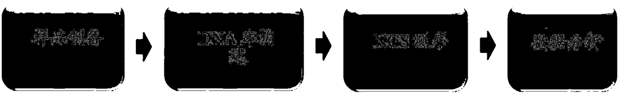
[0094] As shown in Table 2 below, using the same method as in Example 2, 318 lung cancer patients (90 adenocarcinoma, 66 small cell carcinoma, 91 squamous cell carcinoma, 77 other types) and 207 patients were used DNA was extracted from the serum samples of COPD patients, and then PCR was performed on them using 16S rDNA primers to amplify the DNA, followed by sequencing (Illumina MiSeq sequencer). Output the result as a standard flow chart (SFF) file, and use GS FLX software (v2.9) to convert the SFF file into a sequence file (.fasta) and nucleotide quality score file, and then determine the credit rating for reading, And remove those with less than 99% (Phred score The part of the average base call accuracy of the window (29bps) of <20). After deleting low-quality parts, only reads with a length of 300 bp or greater (Sickle version 1.33) were used, and for operational taxon (OTU) ...


PUM


Abstract

The invention relates to a kit and method for the diagnosis of lung cancer in chronic obstructive pulmonary disease (COPD) patients through meta-genome analysis. The invention provides a kit for providing diagnosis information of lung cancer through bacteria meta-genome analysis. More specifically, the invention provides a kit for predicting the risk of lung cancer or diagnosing lung cancer basedon the increasing or decreasing of extracellular vesicles of specific bacteria of a sample of a tested patient through bacterial meta-genome analysis. The extracellular vesicles (EV) secreted from thebacteria present in an environment can be absorbed into the body and directly affect cancer development. Lung cancer is difficult to diagnose before symptoms occur, thereby making effective treatmentdifficult. According to the present invention, the meta-genomes of the extracellular vesicles (EV) present in the human-derived sample are analyzed to predict the risk of lung cancer, thereby predicting and diagnosing the risk groups of the lung cancer to prevent or delay the incidence of lung cancer through appropriate management. The present invention also enables early prediction even after the incidence to lower the incidence rate of the lung cancer and enhance the treatment effects.

Description

[0001] Cross references to related applications [0002] This application claims the priority and rights of Korean Patent Application No. 2016-0179225 filed on December 26, 2016 and Korean Patent Application No. 2017-0080905 filed on June 27, 2017, the disclosure of which is incorporated by reference in its entirety Into this article. Technical field [0003] The present disclosure relates to methods and kits for providing information for diagnosis of lung cancer through metagenomic analysis of bacteria, and more specifically, to metagenomic analysis of bacteria through the use of samples derived from subjects, by analyzing the source The increase or decrease in the content of extracellular vesicles of specific bacteria can be used to predict the risk of lung cancer or to use methods and kits for diagnosing lung cancer. Background technique [0004] Lung cancer is a malignant tumor that originates from the lungs. Despite the development of modern medical science, less than 50% of p...

Claims


Application Information

Patent Timeline
Patent Type & Authority Applications(China)
IPC IPC(8): C12Q1/6886
CPCC12Q1/686C12Q1/6886C12Q1/689C12Q2600/166C12Q2531/113C12Q2600/158C12Q1/6869C12Q2535/122C12Q1/6895C12Q1/6806
Inventor 金润根全成圭
Owner MD HEALTHCARE INC
Why Eureka
Try Eureka