Method and device for segmentation of breast cancer pathologic image

A technology for pathological images and breast cancer, applied in the field of biomedical information, can solve problems such as poor initialization contour results, poor segmentation results, and over-segmentation

Active Publication Date: 2018-08-03
BEIHANG UNIV
View PDF5 Cites 64 Cited by
  • Summary
  • Abstract
  • Description
  • Claims
  • Application Information

AI Technical Summary

Problems solved by technology

However, the Otsu method performs poorly in segmentation in histopathological images, often resulting in poor initial contour results
Although the watershed method improves the segmentation effect compared with the Otsu method, the watershed method is easy to cause over-segmentation. Using the segmentation result of the watershed method to initialize the contour will segment the non-cellular area, resulting in the segmentation of some background area pixels.

Method used


Image

  • Method and device for segmentation of breast cancer pathologic image
  • Method and device for segmentation of breast cancer pathologic image
  • Method and device for segmentation of breast cancer pathologic image

Examples


Embodiment Construction

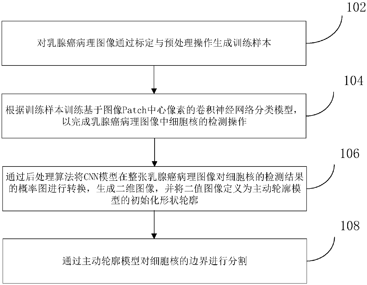
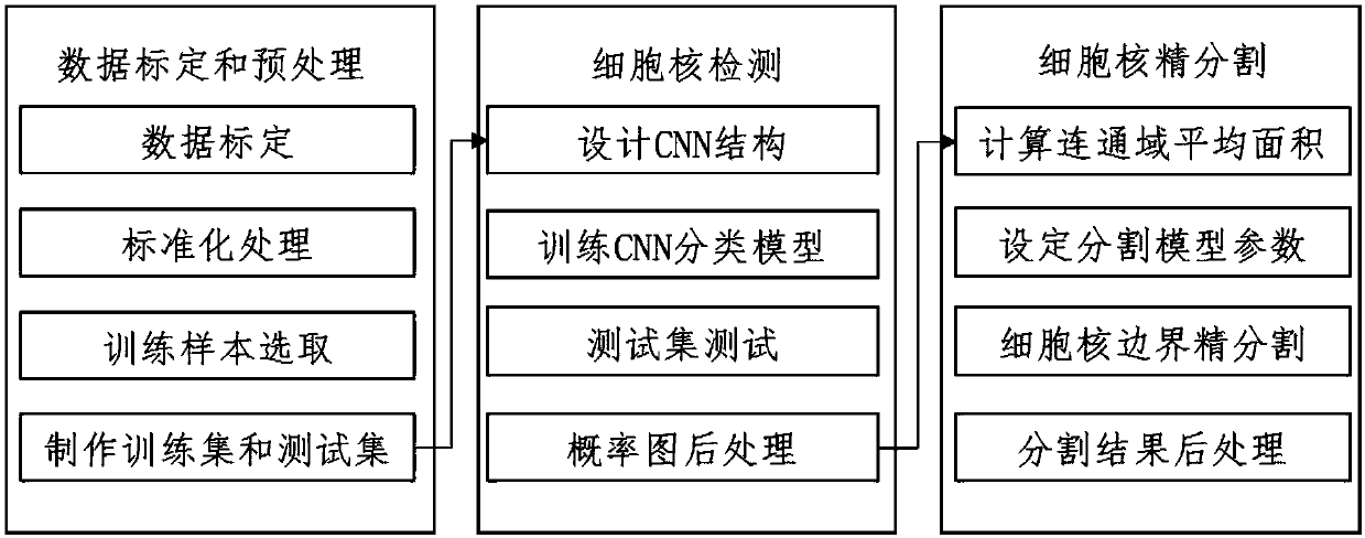
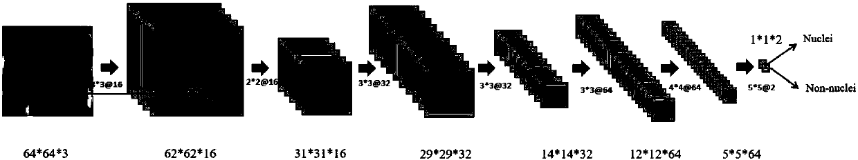
[0033] In order to make the purpose, technical solution and advantages of the present invention clearer, the specific implementation of the method and device for breast cancer pathological image segmentation of the present invention will be further described in detail through the following examples and in conjunction with the accompanying drawings. It should be understood that the specific embodiments described here are only used to explain the present invention, not to limit the present invention.

[0034] The invention relates to the technical field of biomedical information, in particular to the field of pathological image segmentation algorithm research. A segmentation method and device for pathological images of breast cancer is proposed. Specifically, a breast cancer pathological image segmentation method and device based on deep learning and active contour model are provided.

[0035] to combine Figure 1-Figure 5 as shown, figure 1 It is a schematic flowchart of the...


PUM


Abstract

The present invention discloses a method for segmentation of a breast cancer pathologic image. There are three main modules consisting of data preprocessing, cell nucleus detection and cell nucleus boundary refinement segmentation. Pathology experts perform artificial calibration of a cell nucleus boundary, the pathologic image is subjected to standardization processing to eliminate dying difference, a training sample based on a cell nucleus pixel, and a cell nucleus boundary pixel and a background pixel is made to train a convolutional neural network classifier and achieve a classifier basedon a Patch image center pixel. A trained convolutional neural network model is detected on the whole pathologic image to output a probability graph, a postprocessing algorithm is employed to generatea binary image as an initialized shape outline of an active outline model, and the active outline model is employed to perform refinement segmentation of the cell nucleus boundary. The method and thedevice for segmentation of the breast cancer pathologic image are high in segmentation accuracy, and can achieve the algorithm for segmentation of overlapped cells in the breast cancer pathologic image. The present invention further discloses a device for segmentation of a breast cancer pathologic image.

Description

technical field [0001] The invention relates to the technical field of biomedical information, in particular to a segmentation method and device for pathological images of breast cancer. Background technique [0002] In order to solve the practical difficulties encountered in traditional histopathological analysis, digital pathology has developed rapidly in recent years. Digital pathology is a new and rapidly growing field of medical imaging. Tissue slices are digitized after being scanned with ultra-high resolution by the scanner, and then the obtained images are automatically stitched and processed with high precision and multi-view seamlessly by computer, so as to obtain high-quality visualized data for application in various fields of pathology. The application of digital pathology system first started in 1985, and began to be used in the commercial field in the United States in the 1990s. Since 2000, it has gradually replaced traditional microscopes in medical schools....

Claims


Application Information

Patent Timeline
Patent Type & Authority Applications(China)
IPC IPC(8): G06T7/00G06T7/10
CPCG06T7/0012G06T7/10G06T2207/20084G06T2207/20081G06T2207/30068
Inventor 万涛秦曾昌赵磊
Owner BEIHANG UNIV
Why Eureka
Try Eureka