
Fast radiological imaging method, digital radiological imaging device and storage medium

A digital radiography, radiography technology, applied in medical imaging, health care informatics, instruments, etc., can solve the problems of cumbersome, large number of patients, reduced operation efficiency, etc., to improve the degree of automation, improve the efficiency of radiography and users Experience the effect of reducing the number of manual operations

Active Publication Date: 2018-11-02
SHENZHEN MINDRAY BIO MEDICAL ELECTRONICS CO LTD
View PDF8 Cites 4 Cited by
  • Summary
  • Abstract
  • Description
  • Claims
  • Application Information

AI Technical Summary

Problems solved by technology

Using the existing DR operating software to complete each of the above steps requires the operator to perform manual operations, which is very cumbersome
[0004] The characteristic of general radiology examination is a large number of patients, so the operation process of the examination is required to be simplified as much as possible to improve efficiency, and frequent manual operations will undoubtedly greatly reduce the operation efficiency, further reducing the number of patients that digital radiography devices can examine every day

Method used


Image

  • Fast radiological imaging method, digital radiological imaging device and storage medium
  • Fast radiological imaging method, digital radiological imaging device and storage medium
  • Fast radiological imaging method, digital radiological imaging device and storage medium

Examples


Embodiment 1

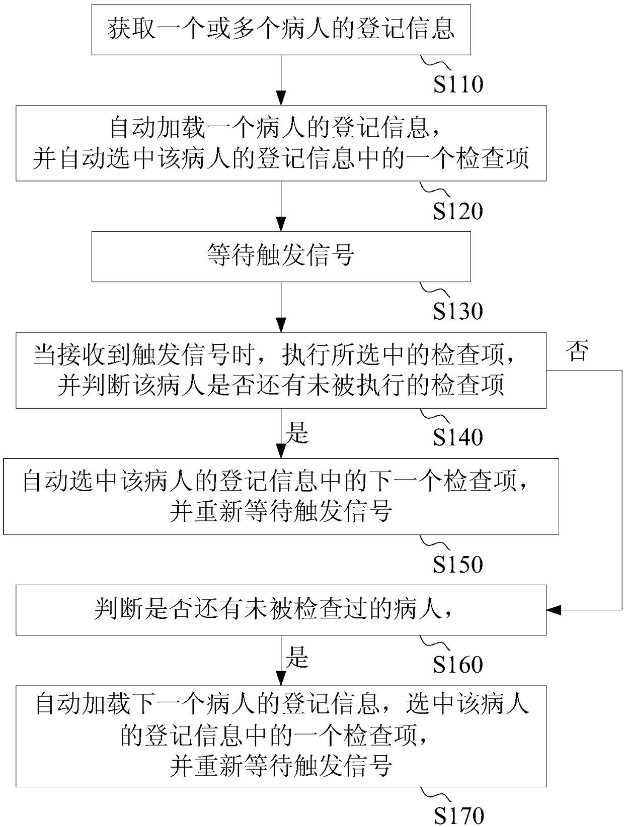
[0061] Please refer to figure 1 , an embodiment discloses a quick inspection method, which includes steps S110-S170.

[0062] Step S110: Obtain the registration information of one or more imaging targets (for example, patients or animals or other objects to be subjected to radiological examination, etc., the following may all take patients as imaging targets for illustration), wherein each patient's The registration information includes one or more imaging items (also referred to as inspection items, which are radiographic imaging items to be performed on the imaging target, which may be described below as inspection items instead of imaging items). In addition to examination items, the patient's registration information may also include some basic information of the patient, such as name, gender, age, assigned examination number, and so on.

[0063] In one embodiment, obtaining the registration information of one or more patients includes at least one of the following method...

Embodiment 2

[0085] Please refer to Figure 4 , this embodiment also provides another rapid radiographic imaging method, which includes steps S210-S240.

[0086] Step S210: storing the registration information of multiple patients, wherein the registration information of each patient includes information of one or more examination items to be performed on this patient. In one embodiment, the patient's registration information can be stored in a self-defined manner, or in the form of a standard examination list, for example, the patient's registration information is stored in the form of Worklist, where Worklist is a DICOM standard examination list.

[0087] In an embodiment, the registration information may include inspection parameters corresponding to each inspection item information. In an embodiment, the examination item information may include information describing the body part to be examined and the patient's position. In addition, the patient's registration information may incl...


PUM


Abstract

The invention discloses a fast radiological imaging method, a digital radiological imaging device and a storage medium. According to the fast radiological imaging method, the digital radiological imaging device and the storage medium, the switching of different imaging items of the same imaging target and / or switching of different imaging targets can be completed by just one action, the number ofmanual operations of an operator is greatly reduced, the automation degree of the workflow is improved, and the imaging efficiency and user experience are significantly improved.

Description

technical field [0001] The invention relates to a fast radiographic imaging method, a digital radiographic imaging device and a storage medium. Background technique [0002] The operation process and operation software of Digital Radiology (DR, Digital Radiology) devices are very complicated. Taking DR operation software as an example, it is usually divided into modules such as patient management, examination management, and image browsing. These functional modules are usually divided into different interfaces. superior. In order to complete the radiographic examination of the patient, the operator usually needs to switch between different interfaces, and also select data and perform operations on different interfaces. In this way, it takes several times to complete a radiographic examination. Interface switching and operation greatly reduces work efficiency. [0003] For example, taking the conventional exposure inspection in radiographic examination as an example, it is ...

Claims


Application Information

Patent Timeline
IPC IPC(8): G16H30/00G16H40/63
CPCG06T11/003
Inventor 张继晔刘彦朋张赞超周华刘兵全
Owner SHENZHEN MINDRAY BIO MEDICAL ELECTRONICS CO LTD
Learn More