A 3D Adversarial Network Based Medical CT Image Segmentation Method

A CT image and network technology, applied in the field of medical image analysis, can solve problems such as instance blurring, achieve the effects of preventing over-fitting, simple method design, and reduced training time

Active Publication Date: 2021-07-09
SHENZHEN INST OF FUTURE MEDIA TECH
View PDF7 Cites 0 Cited by
  • Summary
  • Abstract
  • Description
  • Claims
  • Application Information

AI Technical Summary

Problems solved by technology

Compared with variational self-encoders, GAN does not introduce any deterministic bias (deterministic bias). Variational methods introduce a deterministic bias because they optimize the lower bound of the log-likelihood, rather than the likelihood itself, which leads to VAE Generated instances are blurrier than GAN

Method used


Image

  • A 3D Adversarial Network Based Medical CT Image Segmentation Method
  • A 3D Adversarial Network Based Medical CT Image Segmentation Method
  • A 3D Adversarial Network Based Medical CT Image Segmentation Method

Examples


Embodiment Construction

[0050] The present invention will be further described in detail below in combination with specific embodiments and with reference to the accompanying drawings. It should be emphasized that the following description is only exemplary and not intended to limit the scope of the invention and its application.

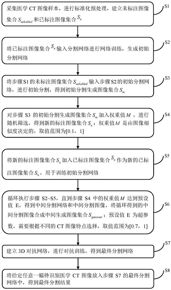
[0051] Whole method step S1~S8 of the present invention please refer to figure 1 . figure 1 It is a flowchart of a 3D confrontation network-based medical CT image segmentation method of the present invention.

[0052] A kind of medical CT image segmentation method based on 3D confrontation network that the present invention proposes, comprises the following steps:

[0053] S1: Collect medical CT image samples, perform standardized preprocessing, and establish an unlabeled image set S unlabled and the set of labeled images S u .

[0054] Perform standardized preprocessing on the collected medical CT image samples. Randomly extract half of the images after standardized...


PUM


Abstract

The invention discloses a medical CT image segmentation method based on a 3D confrontation network, comprising the following steps: collecting medical CT image samples, performing standardized preprocessing; establishing a final segmentation network; putting any given medical CT image to be identified into the final In the segmentation network, the final segmentation result is obtained. In the present invention, the structural form of the 3D confrontation network is designed as a whole, and the segmentation network is used as the generation network G of the 3D confrontation network, and the pre-trained VGG11 is used as the discrimination network D of the 3D confrontation network. The invention uses the confrontation network to increase the labeling data, and through similarity calculation and cyclic random screening, the credibility of the image is enhanced to generate high-quality labeling image collections in batches, and finally the segmentation is updated through the confrontation training of the discriminant network D and the segmentation network Network parameters and discriminant network D parameters, so as to optimize the segmentation network and improve the segmentation accuracy. The method of the invention is simple in design and easy to implement.

Description

technical field [0001] The invention belongs to the field of medical image analysis, and relates to a method for segmenting regions of interest in medical CT images. Background technique [0002] With the development of medical imaging technology, medical imaging examination is becoming more and more important in clinical diagnosis. Computed Tomography (CT) is the most commonly used method among many medical imaging techniques. At present, the diagnosis of medical images is mainly completed by manual reading by doctors. Therefore, differences in the personal experience and knowledge level of doctors will affect the accuracy of diagnosis. For example, a CT image of the lungs is a screenshot of a cross-section of the entire chest, and often contains tissue information of many other organs, which seriously interferes with the doctor's diagnostic work. Therefore, it is very necessary to use computer technology to process CT images. How to accurately segment the doctor's regi...

Claims


Application Information

Patent Timeline
Patent Type & Authority Patents(China)
IPC IPC(8): G06T7/10
CPCG06T2207/10081G06T2207/20081G06T2207/20084G06T7/10
Inventor 张颖洪晓东王好谦
Owner SHENZHEN INST OF FUTURE MEDIA TECH
Why Eureka
Try Eureka