Three-dimensional breast ultrasound scanning method and ultrasound scanning system

A technology for ultrasound scanning and breast cancer, applied in the field of ultrasound scanning, can solve the problems of not covering the nipple, the relative position of the lesion is not accurate, etc., and achieve the effect of improving the accuracy rate

Active Publication Date: 2019-01-11
ZHEJIANG SHENBO MEDICAL TECH CO LTD
View PDF12 Cites 6 Cited by
  • Summary
  • Abstract
  • Description
  • Claims
  • Application Information

AI Technical Summary

Problems solved by technology

[0003] The problem with the existing technology is that if the ultrasound scan image is on both sides, the nipple may not be covered in the scan image. At this time, the approximate po...

Method used


Image

  • Three-dimensional breast ultrasound scanning method and ultrasound scanning system
  • Three-dimensional breast ultrasound scanning method and ultrasound scanning system
  • Three-dimensional breast ultrasound scanning method and ultrasound scanning system

Examples


Embodiment Construction

[0037] In the following description, many technical details are proposed for the reader to better understand this application. However, those of ordinary skill in the art can understand that even without these technical details and various changes and modifications based on the following embodiments, the technical solution claimed in this application can be realized.

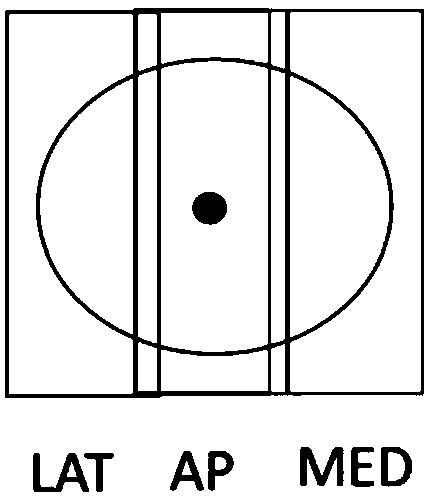
[0038] Explanation of some concepts:

[0039] AP: Physical location of breast center

[0040] MED: physical location of the inside of the breast (near the central axis)

[0041] LAT: Physical location on the outside of the breast (near the arm)

[0042] In order to make the objectives, technical solutions, and advantages of the present application clearer, the implementation manners of the present application will be described in further detail below in conjunction with the accompanying drawings.

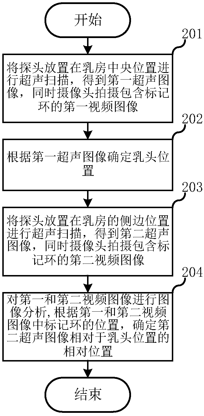
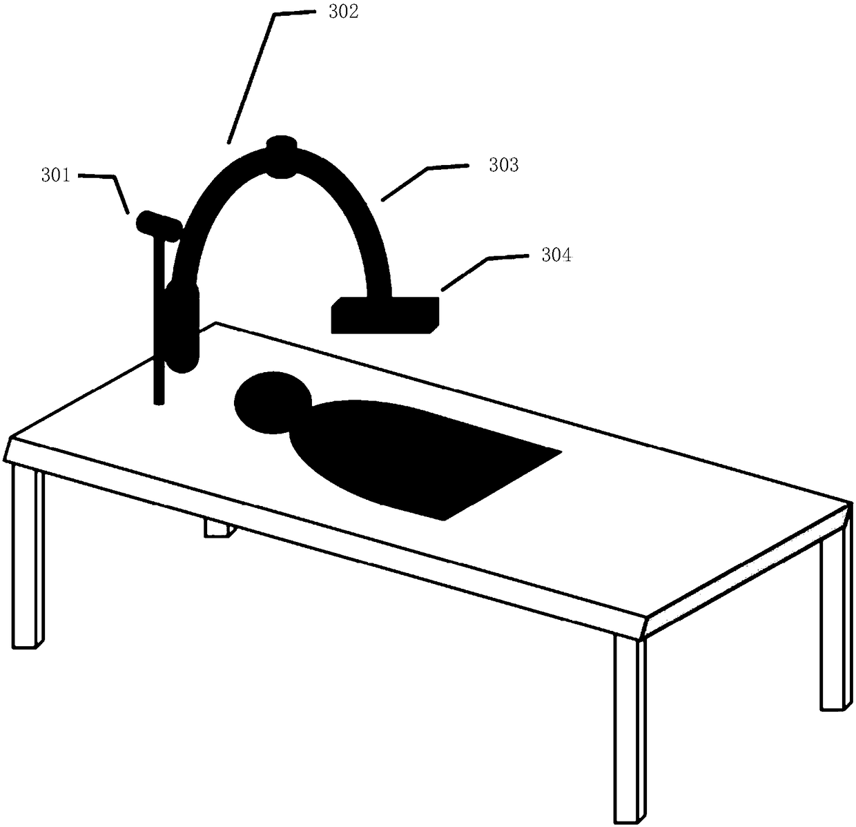
[0043] The first embodiment of the present invention relates to a method for performing three-dimensional breast ultrasound s...


PUM


Abstract

The present application relates to the field of ultrasound scanning, and discloses a three-dimensional breast ultrasound scanning method and an ultrasound scanning system, being able to accurately andautomatically determine the position of the nipple with respect to the lateral ultrasound image. In the method, putting a probe at a central position of the breast for ultrasonic scanning to obtain afirst ultrasonic image while a camera captures a first video image including a marker ring, wherein the first ultrasonic image covers a nipple position; determining a nipple position according to thefirst ultrasound image; placing a probe at a side position of the breast for ultrasonic scanning to obtain a second ultrasonic image, and simultaneously a camera captures a second video image including a marker ring; and performing image analysis on the first and second video images to determine a relative position of the second ultrasound image with respect to the nipple position based on the position of the marker ring in the first and second video images.

Description

Technical field [0001] This application relates to the field of ultrasound scanning, in particular to breast ultrasound scanning technology. Background technique [0002] At present, when the automatic breast ultrasound scanning system is used to scan the breast, the three-dimensional image of an automatic scan can cover a part of the breast. In diagnosis or screening, in order to cover the entire breast, the operator usually needs to place the ultrasound probes in three positions of the breast to obtain three three-dimensional images to cover the entire breast area. These three positions are usually AP (central breast), MED (inner breast), LAT (outer breast), such as figure 1 Shown are the three positions of the right breast. The obtained three-dimensional image will also be marked as the corresponding location to facilitate the doctor to read the film. And this marking behavior is usually manually input by the operator in the scanning workstation. In the acquired image, due ...

Claims


Application Information

Patent Timeline
IPC IPC(8): A61B8/08
CPCA61B8/0825A61B8/483A61B8/5238
Inventor 朱轲梁浈檀韬
Owner ZHEJIANG SHENBO MEDICAL TECH CO LTD
Why Eureka
Try Eureka