A lung parenchyma CT image segmentation method based on a deep convolutional neural network

A neural network and deep convolution technology, applied in the field of medical image processing and deep learning, can solve the problems of difficult segmentation of lungs, unsatisfactory segmentation effect, and limited practical application, etc., to improve accuracy, fast algorithm speed, and reduce calculation volume effect

Pending Publication Date: 2019-04-16
TIANJIN POLYTECHNIC UNIV
View PDF8 Cites 18 Cited by
  • Summary
  • Abstract
  • Description
  • Claims
  • Application Information

AI Technical Summary

Problems solved by technology

The contour segmentation method cannot deal with images with uneven gray levels very well, which limits its practical application in medical images to a certain extent; in addition, the three existing lung parenchyma segmentation methods

Method used


Image

  • A lung parenchyma CT image segmentation method based on a deep convolutional neural network
  • A lung parenchyma CT image segmentation method based on a deep convolutional neural network
  • A lung parenchyma CT image segmentation method based on a deep convolutional neural network

Examples


Embodiment Construction

[0018] The detailed description is as follows with reference to the attached preferred embodiments, and the described preferred embodiments are only used to illustrate the technical solution of the present invention, not to limit the present invention.

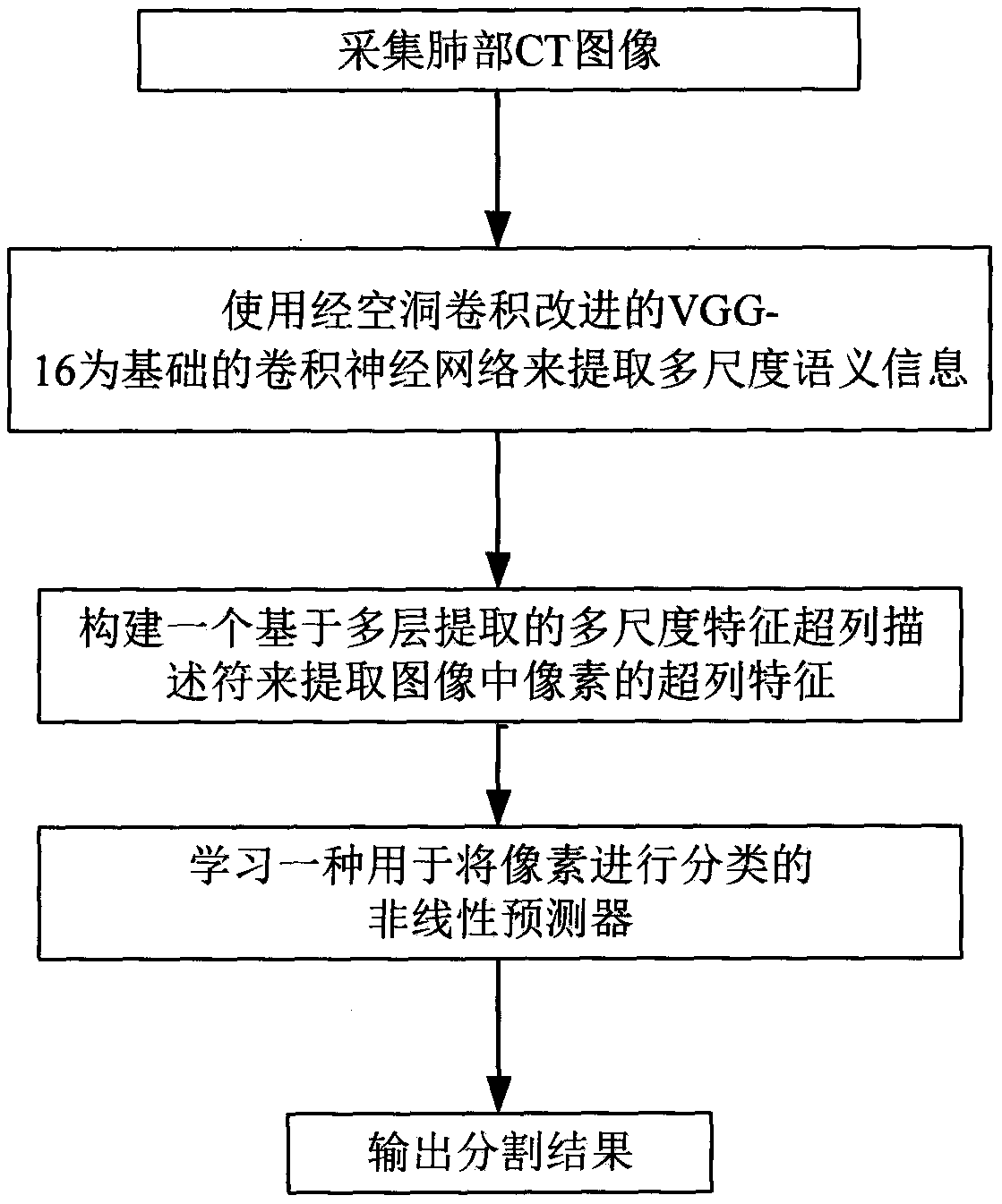
[0019] figure 1 A flow chart of the lung parenchyma CT image segmentation method based on the deep convolutional neural network according to the present invention is given. Such as figure 1 Shown, according to the lung parenchyma CT image segmentation method based on deep convolutional neural network of the present invention comprises:

[0020] Step 1: collecting lung CT images;

[0021] Step 2: Use the VGG-16-based convolutional neural network improved by dilated convolution to extract multi-scale semantic information;

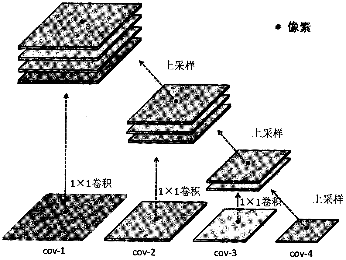
[0022] Step 3: Construct a multi-scale feature hypercolumn descriptor based on multi-layer extraction to extract hypercolumn features of pixels in the image;

[0023] Step 4: Learn a non-linear predictor ...


PUM


Abstract

The invention designs a lung parenchyma CT image segmentation method based on a deep convolutional neural network. The method comprises the steps that 1, using The VGG-16 network improved by cavity convolution to extract the convolution characteristics of lung parenchyma in lung CT images;; 2, constructing a multi-scale feature supercolumn descriptor based on multi-layer extraction; And 3, learning a nonlinear predictor for classifying pixels. Results show that the method can realize accurate lung parenchyma image segmentation, compared with the prior art, the method has the advantages that the segmentation accuracy and speed are improved, and the problems of low manual segmentation efficiency, low accuracy, excessive consumption of manpower resources and the like of the lung parenchyma image are successfully solved.

Description

technical field [0001] The invention relates to a lung parenchyma CT image segmentation method based on a convolutional neural network, which is superior to the prior art in terms of segmentation efficiency, robustness and accuracy, has good segmentation performance, and belongs to the field of medical image processing and deep learning . Background technique [0002] According to statistics, the incidence of lung cancer in China is increasing at a rate of 26.9% every year. By 2025, the number of people dying of lung cancer in China alone will be close to 1 million per year. Studies have shown that early detection and early treatment of lung cancer can effectively improve the survival rate of lung cancer patients: the 5-year survival rate increased from 14% to 49%. CT imaging is one of the effective ways to help doctors diagnose lung diseases. In order to reduce the workload of doctors and detect lung diseases faster and more accurately, it is of great significance to appl...

Claims


Application Information

Patent Timeline
IPC IPC(8): G06T7/10G06N3/04
CPCG06T7/10G06T2207/30064G06T2207/30101G06T2207/20081G06T2207/10081G06N3/045
Inventor 耿磊刘博文肖志涛张芳吴骏张思奇刘彦北
Owner TIANJIN POLYTECHNIC UNIV
Why Eureka
Try Eureka