A Pulmonary Nodule Segmentation Method Based on 2D Convolutional Neural Network

A two-dimensional convolution, neural network technology, applied in image analysis, image enhancement, instrumentation, etc., can solve the problem of not well adapted to the heterogeneity of pulmonary nodules

Active Publication Date: 2020-10-30
HUAZHONG UNIV OF SCI & TECH
View PDF5 Cites 0 Cited by
  • Summary
  • Abstract
  • Description
  • Claims
  • Application Information

AI Technical Summary

Problems solved by technology

[0005] In view of the defects of the prior art, the purpose of the present invention is to solve the technical problem that none of the prior art can well adapt to the heterogeneity of pulmonary nodules, especially the segmentation of pulmonary nodules with smaller sizes

Method used


Image

  • A Pulmonary Nodule Segmentation Method Based on 2D Convolutional Neural Network
  • A Pulmonary Nodule Segmentation Method Based on 2D Convolutional Neural Network
  • A Pulmonary Nodule Segmentation Method Based on 2D Convolutional Neural Network

Examples


Embodiment Construction

[0041] In order to make the object, technical solution and advantages of the present invention clearer, the present invention will be further described in detail below in conjunction with the accompanying drawings and embodiments. It should be understood that the specific embodiments described here are only used to explain the present invention, not to limit the present invention.
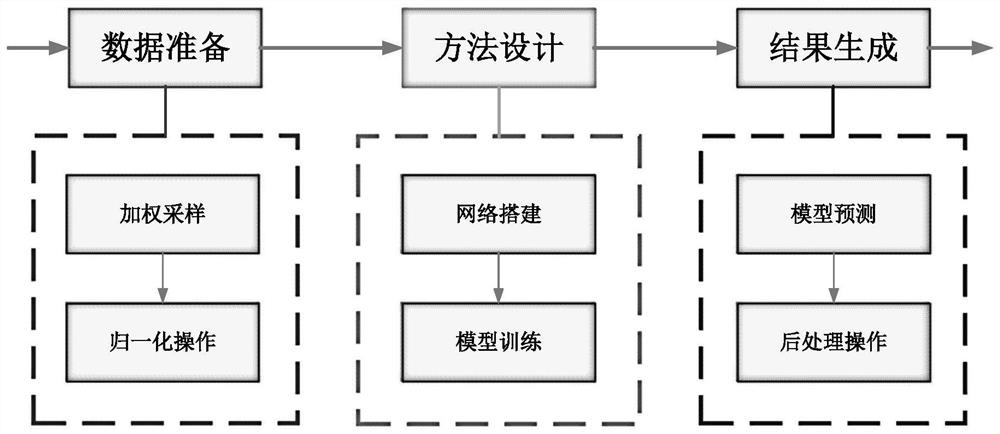
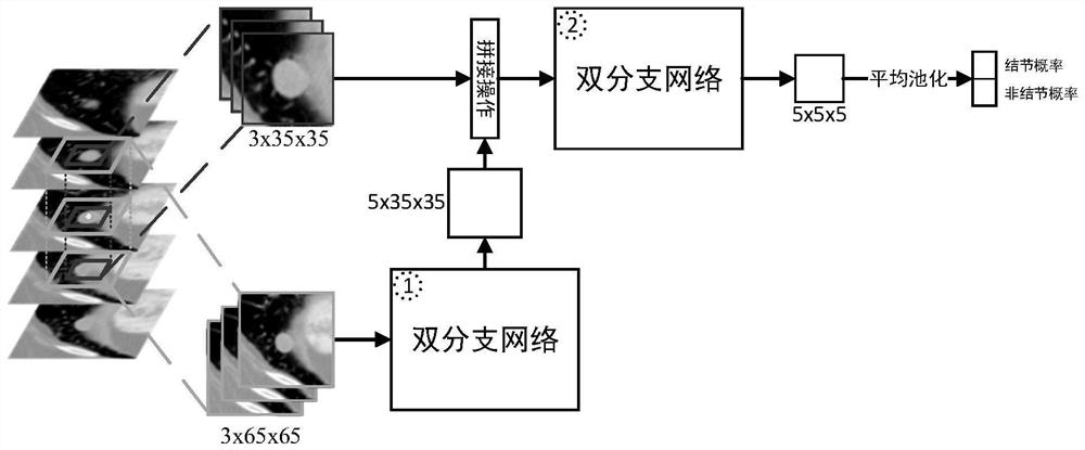
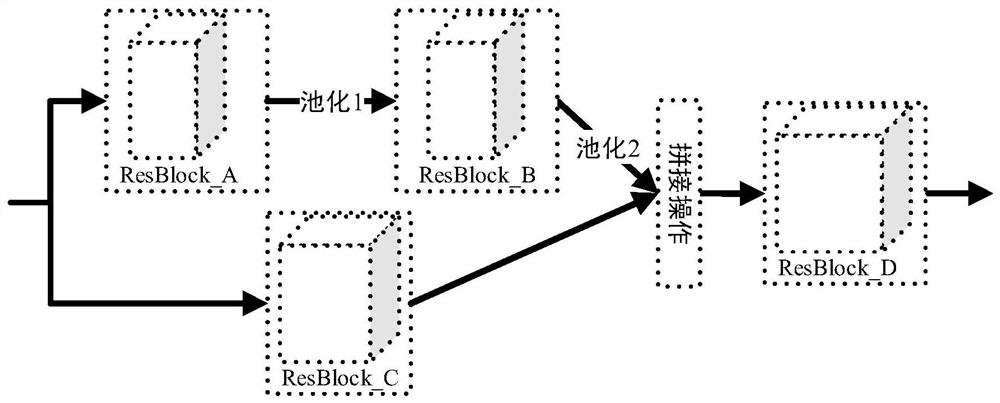
[0042] The overall idea of ​​the present invention is to obtain multi-view and multi-scale features of different pulmonary nodules in CT images in a cascade manner, and use a residual block-based dual-branch network to extract local detail features of pulmonary nodules and the rich context around them information. In addition, the present invention also uses an edge-based weighted sampling strategy to facilitate model training to improve the generalization ability of the model as much as possible.

[0043] Such as figure 1 Shown, a lung nodule segmentation method based on two-dimensional convolut...


PUM


Abstract

The invention discloses a pulmonary nodule segmentation method based on a two-dimensional convolutional neural network. The pulmonary nodule segmentation method comprises the following steps: samplingpulmonary nodule positive and negative samples based on a weighted sampling strategy; training a two-dimensional convolutional neural network model according to the sampled data to obtain a trained two-dimensional convolutional neural network model; and predicting each voxel of the sample to be segmented by using the trained two-dimensional convolutional neural network model to obtain a pulmonarynodule segmentation result. When the sampling weight of non-pulmonary nodule voxels in a CT image is calculated, the grayscale information of non-pulmonary nodule tissues is considered, so that advanced features except the grayscale features are mined, and the heterogeneous property of pulmonary nodules is adapted; Fully sampling pulmonary nodules with different sizes by taking pulmonary nodule edge voxels as references; Local texture information and context information of the pulmonary nodules can be extracted based on the double-branch cascade network of the residual block; Through cascadeconnection of the two image blocks with different scales, segmentation of pulmonary nodules with small sizes is realized.

Description

technical field [0001] The invention belongs to the technical field of image segmentation, and more specifically relates to a pulmonary nodule segmentation method based on a two-dimensional convolutional neural network. Background technique [0002] Pulmonary nodules are a precursor to lung cancer, but the presence of lung nodules does not necessarily mean cancer, that is, lung nodule information is an intermediate result. Accurate segmentation of pulmonary nodules is a key step in computer-aided diagnosis of early lung cancer based on CT images. Whether pulmonary nodules can be accurately segmented from CT images will ultimately affect the performance of computer-aided diagnosis systems. [0003] In the prior art, pulmonary nodule segmentation methods mainly include methods based on morphology and region growth. However, there are problems in robustness, especially for the segmentation of pulmonary nodules adhered to the lung wall. Methods based on deep learning, such as t...

Claims


Application Information

Patent Timeline
Patent Type & Authority Patents(China)
IPC IPC(8): G06T7/12
CPCG06T2207/10081G06T2207/20081G06T2207/20084G06T2207/30064G06T7/12
Inventor 刘宏曹海潮马光志宋恩民刘腾营刘磊刘楚华金勇庄宇舟金人超许向阳
Owner HUAZHONG UNIV OF SCI & TECH
Why Eureka
Try Eureka