A lung anatomy location positioning algorithm based on a deep learning technology

A deep learning and localization algorithm technology, applied in computing, image data processing, instruments, etc., can solve problems such as difficulty in extracting lung fissures, affecting the segmentation effect, and incomplete lung fissures, avoiding the risk of overfitting and ensuring generalization. Ability to ensure the effect of segmentation accuracy

Pending Publication Date: 2019-06-14
成都蓝景信息技术有限公司
View PDF12 Cites 11 Cited by
  • Summary
  • Abstract
  • Description
  • Claims
  • Application Information

AI Technical Summary

Problems solved by technology

[0005] (2) The amount of calculation is large. According to the process described in (1), a large amount of calculation is required, especially when segmenting the trachea and blood vessels, the region growth algorithm is often used. In addition, the Hessian matrix of the image needs to be calculated when the lung fissure is extracted. , and then calculate the eigenvalues ​​and eigenvectors of the matrix, and the amount of CT image information, the above calculation is undoubtedly an extremely time-consuming process;
[0006] (3) The segmentation effect is not good. First, when extracting the lung fissure, other similar graphic structures are extracted, so secondary extraction is often required. Even so, the extracted lung fissure will be broken or interfered by other tissues The problem
In addition, lung fissures are not obvious, fissures are incomplete, or even fissures disappear in lesioned lung CT and non-high-definition CT, which will cause great difficulties in the extraction of lung fissures. Region discontinuity or region error occurs
[0007] (4) The generalization ability is poor. During the segmentation process, it is necessary to set seed points, thresholds, etc. The setting and selection of these values ​​will greatly affect the final segmentation effect
Coupled with the individual differences and geometric diversity of CT images, it is difficult for these methods to meet actual needs.

Method used


Image

  • A lung anatomy location positioning algorithm based on a deep learning technology
  • A lung anatomy location positioning algorithm based on a deep learning technology

Examples


Embodiment Construction

[0020] The following will be combined with Figure 1-Figure 2 The present invention is described in detail, and the technical solutions in the embodiments of the present invention are clearly and completely described. Apparently, the described embodiments are only some of the embodiments of the present invention, not all of them. Based on the embodiments of the present invention, all other embodiments obtained by persons of ordinary skill in the art without making creative efforts belong to the protection scope of the present invention.

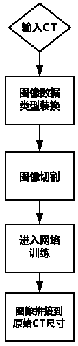
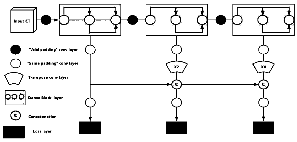
[0021] The present invention provides a lung anatomical position positioning algorithm based on deep learning technology through improvement; this patent proposes an end-to-end deep learning network, which can accurately and quickly divide lung CT, and the process is simple, roughly as follows :

[0022] (1) Data type conversion is performed on the input CT, and the pixel value of the image is converted from a 32-bit floating-point number ty...


PUM


Abstract

The invention discloses a lung anatomy position positioning algorithm based on a deep learning technology, which can accurately and quickly divide lung CT, and can simply, quickly and accurately realize automatic segmentation of lung lobes based on lung CT images, thereby realizing the anatomy position positioning of lung lesions. Compared with a traditional segmentation method, the method has theoutstanding advantages that (1) the process is simple, and the end-to-end segmentation mode does not need to pay attention to other processes; (2) the multi-stage and multi-output network architecture controls the network in different stages, so that the segmentation effect is better, and the segmentation precision can be ensured to the maximum extent through a semantic-based segmentation mode; and (3) the generalization ability is strong, and the data in the training process is enhanced, so that the model can learn different and diverse data, namely, the generalization ability of the segmentation model is ensured, meanwhile, the risk of over-fitting is also avoided to a certain extent, and the geometric deformation and illumination influence of CT (computed tomography) are insensitive when lung lobe division is performed on different CT.

Description

technical field [0001] The present invention relates to a lung anatomical position positioning algorithm, specifically a lung anatomical position positioning algorithm based on deep learning technology. Background technique [0002] The lung is clinically divided into five lobes, the left lung is divided into upper and lower lobes, and the right lung is divided into upper, middle and lower lobes, which is of great significance in medicine. [0003] The main idea of ​​the current lung lobe segmentation algorithm is to extract the "lung fissure" first, and then realize the regional segmentation of the lung lobe according to the lung fissure. These methods typically have the following deficiencies: [0004] (1) The process of the method is complicated. This type of method first needs to segment the lung body, trachea, blood vessels, etc., and remove the interference of these tissues. Then, according to computer graphics, the lung fissures are extracted, and finally, segmentat...

Claims


Application Information

Patent Timeline
Patent Type & Authority Applications(China)
IPC IPC(8): G06T7/10G06T3/40
Inventor 赵继帆
Owner 成都蓝景信息技术有限公司
Why Eureka
Try Eureka