
Automatic right ventricle segmentation method based on deep learnin

An automatic segmentation and deep learning technology, applied in the field of medical image processing, can solve the problems of increased segmentation time, poor performance, and large subjective influence

Inactive Publication Date: 2019-08-13
UNIV OF SHANGHAI FOR SCI & TECH
View PDF4 Cites 22 Cited by
  • Summary
  • Abstract
  • Description
  • Claims
  • Application Information

AI Technical Summary

Problems solved by technology

Commonly used right ventricle segmentation methods include active contour model, level set, graph cut, clustering and multi-atlas segmentation, etc. These methods have shown good results on limited data sets, but often in databases other than training data Poor performance, requiring manual intervention, resulting in increased segmentation time and high subjective impact

Method used


Image

  • Automatic right ventricle segmentation method based on deep learnin
  • Automatic right ventricle segmentation method based on deep learnin
  • Automatic right ventricle segmentation method based on deep learnin

Examples


Embodiment Construction

[0030] Below in conjunction with accompanying drawing, further elaborate the present invention. It should be understood that these examples are only used to illustrate the present invention and are not intended to limit the scope of the present invention. In addition, it should be understood that after reading the teachings of the present invention, those skilled in the art can make various changes or modifications to the present invention, and these equivalent forms also fall within the scope defined by the appended claims of the present application.

[0031] A method for automatically segmenting the right ventricle based on deep learning disclosed in this embodiment includes the following steps:

[0032] (1) Data preprocessing

[0033] The present invention retrospectively analyzes cardiac cine magnetic resonance images (1.5T GE magnetic resonance imaging system). In this embodiment, 844 pieces of MRI image data of 61 patients are included, including 22 males and 39 females...


PUM


Abstract

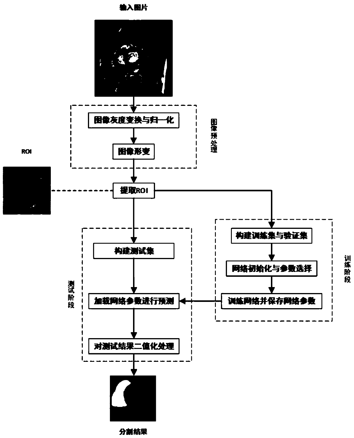
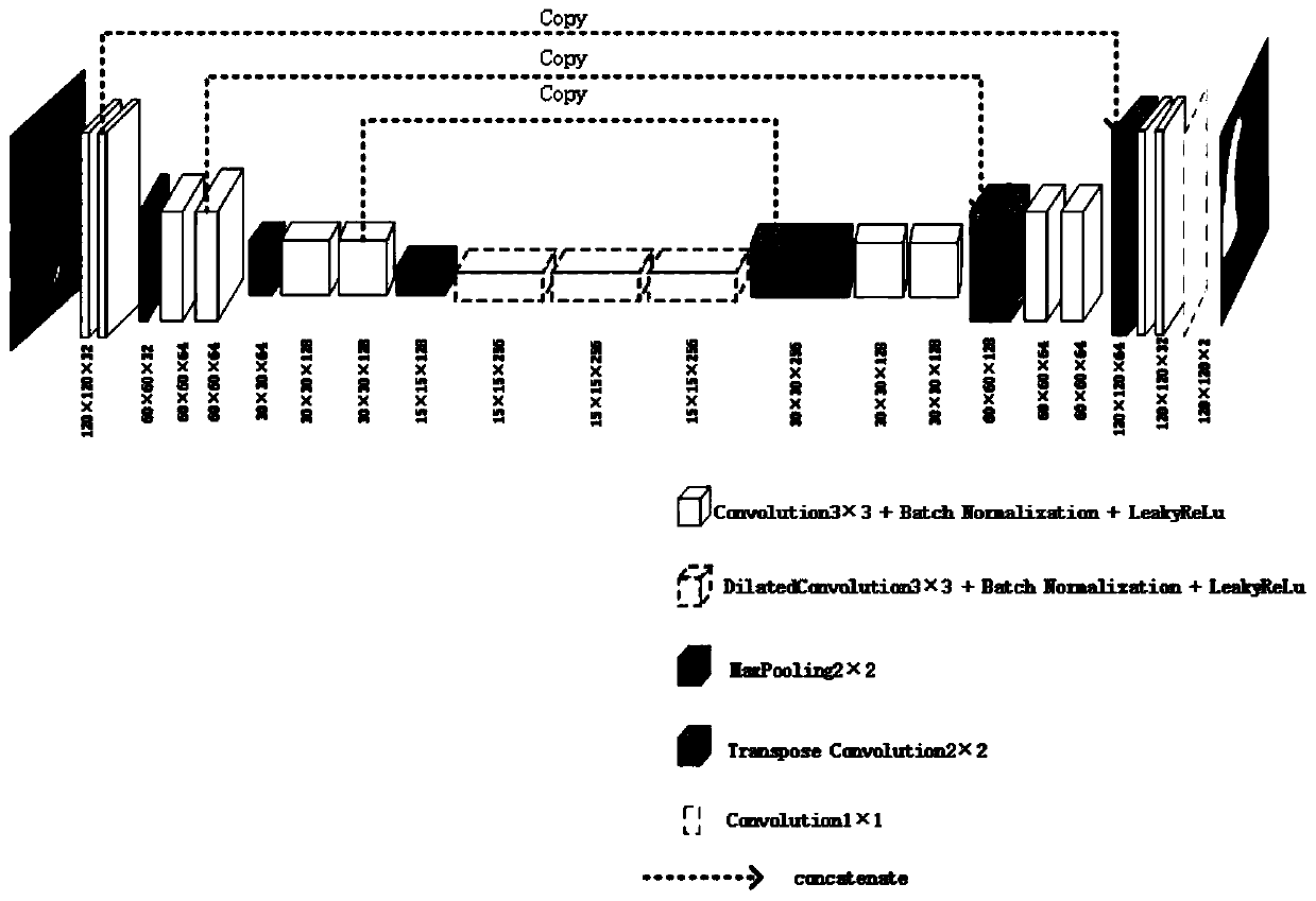
The invention provides an automatic right ventricle segmentation method based on deep learning, and the method is characterized in that the method comprises the following steps: carrying out the preprocessing of all collected heart magnetic resonance film short-axis images; extracting the ROI of the region of interest; expanding the data set; constructing a U-shaped network model in a keras library of deep learning, and training the U-shaped network model; and performing result prediction by using the trained U-shaped network model. The invention provides a full-automatic right ventricle segmentation method based on deep learning. Firstly, the influence of surrounding tissues on a segmentation result is reduced by automatically identifying a ventricle region, and an original U-shaped network is improved, so that accurate and full-automatic segmentation of a right ventricle is realized, and a basis is provided for further functional analysis and disease diagnosis of the heart.

Description

technical field [0001] The invention relates to the field of medical image processing, in particular to an automatic right ventricle segmentation method based on deep learning. Background technique [0002] With the continuous advancement of medical image processing technology, medical image segmentation, especially the segmentation of important organs, is the basis of computer-aided diagnosis and treatment. Magnetic resonance imaging (Magnetic Resonance Imaging) is a standard technique widely used in medical diagnosis. In cardiac magnetic resonance cine images, accurate segmentation of the right ventricle can help people effectively calculate the parameters of the ventricle at the end of systole and end of diastole, stroke volume, and ejection fraction, so as to further analyze the function of the heart and diagnose diseases. [0003] The left ventricle has a regular shape and is surrounded by thicker myocardium. Its segmentation task is relatively easy and has been studie...

Claims


Application Information

Patent Timeline
Patent Type & Authority Applications(China)
IPC IPC(8): G06T7/11G06T7/143G06T7/66
CPCG06T7/11G06T7/143G06T7/66G06T2207/10088G06T2207/20061G06T2207/20076G06T2207/20224G06T2207/30048G06T2207/20081G06T2207/20084
Inventor 刘鹏王丽嘉
Owner UNIV OF SHANGHAI FOR SCI & TECH
Learn More