A nanoscale quantum three-dimensional thermal imaging system for tumor cells

A tumor cell and imaging system technology, applied in the field of tumor cell nano-scale quantum three-dimensional thermal imaging system, can solve the problems of losing temperature information, unfavorable temperature field analysis of tumor cells, etc., and achieve the effect of non-destructive detection

Active Publication Date: 2020-04-10
XI AN JIAOTONG UNIV
View PDF5 Cites 0 Cited by
  • Summary
  • Abstract
  • Description
  • Claims
  • Application Information

AI Technical Summary

Problems solved by technology

Today's cell thermal imaging methods are mostly limited to the average temperature of the plane, which loses the axial temperature information, which is not conducive to the analysis of the temperature field of the biological micro-nano structure of tumor cells

Method used


Image

  • A nanoscale quantum three-dimensional thermal imaging system for tumor cells

Examples


Embodiment Construction

[0016] The present invention will be described in detail below in conjunction with the accompanying drawings and embodiments.

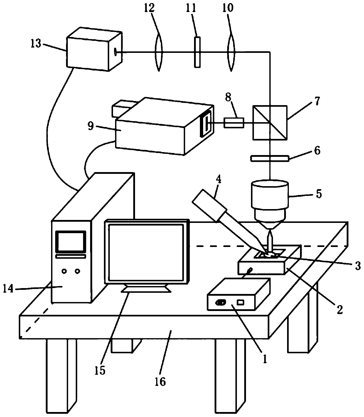
[0017] Such as figure 1 As shown, a tumor cell nanoscale quantum three-dimensional thermal imaging system includes a nanopositioning platform 2, the control input of the nanopositioning platform 2 is connected to the output of the controller 1, and the controller 1 realizes the control of the nanopositioning platform through pre-programmed input instructions. 2. The control of the movement process. The tumor cell sample 3 is placed on the nano-positioning platform 2. The nano-positioning platform 2 is used to move the tumor cell sample 3 at the nanoscale in the axial direction, thereby realizing layered thermal imaging of the tumor cell sample 3. Tumor The top of the cell sample 3 is provided with an excitation light source 4 and a microscope objective lens 5, the output of the microscope objective lens 5 is connected through the input of the optical ...


PUM


Abstract

A tumor cell nano-scale quantum three-dimensional thermal imaging system comprises a nanometer positioning platform connected to output of a controller. A tumor cell sample is placed on the nanometer positioning platform, an excitation light source and a microscope objective lens are arranged above the sample, the output of the microscope objective lens is connected to a beam splitter mirror through a light filter, and light is split into reflected light and transmitted light by the beam splitter mirror. The reflected light is connected to input of a spectrograph through a tube mirror, the output of the spectrograph is connected with first input of a work station, and the tube mirror and the spectrograph form a quantum temperature measuring light path. The transmitted light is connected to the input of an EMCCD detector through a first convex lens, a phase modulation sheet and a second convex lens, and the output of the EMCCD detector is connected to second input of the work station. The first convex lens, the phase modulation sheet, the second convex lens and the EMCCD detector form a three-dimensional positioning light path, and the output of the work station and the input of a display are connected. In the invention, real-time detection of a temperature field in a tumor cell is realized in rapid and non-destructive modes, and a three-dimensional temperature field of the tumor cell is obtained.

Description

technical field [0001] The invention relates to the technical field of biological thermal imaging, in particular to a nanoscale quantum three-dimensional thermal imaging system for tumor cells. Background technique [0002] The study of tumor cell lesion mechanism is a hotspot in the field of biomedicine, and the micro-nano scale heat generation and transfer under unconventional conditions is directly related to tumor cell metabolism and lesion. Due to the heat sensitivity of tumor cells, temperature itself is a key factor in the pathological research and comprehensive treatment of tumor cells. For example, high fever can reduce the metabolism of tumor cells, enhance the killing effect of radiation and improve the chemotherapy effect of drugs. The research on exploring tumor cell therapy methods is also going deeper into the thermal analysis of biomolecules, such as the thermal tolerance of tumor cells related to heat shock protein (HSP), and the immune response initiated by...

Claims


Application Information

Patent Timeline
Patent Type & Authority Patents(China)
IPC IPC(8): G01K11/00
CPCG01K11/00
Inventor 杨军凌正杜汉良李本强梅雪松朱绍冲
Owner XI AN JIAOTONG UNIV
Why Eureka
Try Eureka