Image enhancement method, device and storage medium suitable for endoscope

A technology of image enhancement and endoscopy, which is applied in the field of medical devices, can solve the problems of ineffective enhancement of texture and details, low enhancement effect and low visibility of details, so as to improve the enhancement effect and visibility of details, reduce the processing range, and improve grayscale. The effect of degree contrast

Active Publication Date: 2022-05-27
YUXIN TECH HUIZHOU
View PDF7 Cites 0 Cited by
  • Summary
  • Abstract
  • Description
  • Claims
  • Application Information

AI Technical Summary

Problems solved by technology

However, the inventor found in the process of implementing the present invention that often when a doctor observes a medical image, what he observes is the lesion area of ​​the image, but because the global enhancement method cannot effectively enhance the local texture and details, it can only enhance Global contrast, which enhances the visual effect globally, thus resulting in less enhancement and less detail visibility in the area to be observed

Method used


Image

  • Image enhancement method, device and storage medium suitable for endoscope
  • Image enhancement method, device and storage medium suitable for endoscope
  • Image enhancement method, device and storage medium suitable for endoscope

Examples


Embodiment Construction

[0044] The technical solutions in the embodiments of the present invention will be clearly and completely described below with reference to the accompanying drawings in the embodiments of the present invention. Obviously, the described embodiments are only a part of the embodiments of the present invention, rather than all the embodiments. Based on the embodiments of the present invention, all other embodiments obtained by persons of ordinary skill in the art without creative efforts shall fall within the protection scope of the present invention.

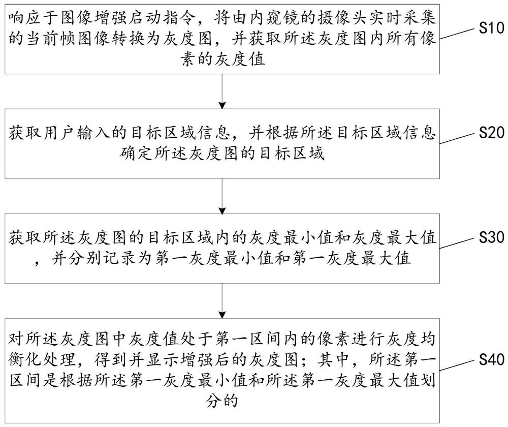
[0045] see figure 1 , is a schematic flowchart of an embodiment of the image enhancement method suitable for an endoscope provided by the present invention.

[0046] An embodiment of the present invention provides an image enhancement method suitable for an endoscope, including steps S10 to S40, and the details are as follows:

[0047]S10. In response to the image enhancement start instruction, convert the current frame image capt...


PUM


Abstract

The invention discloses an image enhancement method, device and storage medium suitable for an endoscope. The method includes the steps of: converting the current frame image collected in real time by a camera of the endoscope into a grayscale image in response to an image enhancement start instruction , and obtain the grayscale values ​​of all pixels in the grayscale image; obtain the target area information input by the user, and determine the target area of ​​the grayscale image according to the target area information; obtain the target area of ​​the grayscale image The gray minimum value and gray maximum value in the gray scale are recorded as the first gray scale minimum value and the first gray scale maximum value respectively; Perform grayscale equalization processing on pixels in the first interval divided by the first grayscale maximum value to obtain and display an enhanced grayscale image. The invention can improve the enhancement effect and detail visibility of the target area in the video image collected by the endoscope.

Description

technical field [0001] The present invention relates to the technical field of medical devices, and in particular, to an image enhancement method, a device and a storage medium suitable for an endoscope. Background technique [0002] Endoscope is a commonly used medical device, which integrates traditional optics, ergonomics, precision machinery, modern electronics, mathematics and software. It is widely used in non-invasive and minimally invasive surgery. When using an endoscope for the diagnosis and treatment of diseases that do not have strict color requirements but high detail display requirements, due to the influence of interfering gray scales, the details displayed by the images collected by the endoscope are not ideal, and the usually obtained medical images may There are phenomena such as unclear details and low contrast, which will affect the safety and effectiveness of diagnosis and treatment. Therefore, making the images collected by the endoscope show clearer t...

Claims


Application Information

Patent Timeline
Patent Type & Authority Patents(China)
IPC IPC(8): G06T5/00
CPCG06T5/009G06T2207/10068
Inventor 宋翀绂孙光宇
Owner YUXIN TECH HUIZHOU
Why Eureka
Try Eureka