
Esophageal intubation detection system for digestive department and image enhancement method

A detection system and image enhancement technology, applied in the field of medical devices, can solve problems such as inconspicuous response, difficult cost problem, and consumption of detection equipment

Pending Publication Date: 2021-05-18
JILIN UNIV FIRST HOSPITAL
View PDF0 Cites 0 Cited by
  • Summary
  • Abstract
  • Description
  • Claims
  • Application Information

AI Technical Summary

Problems solved by technology

[0004] One is a syringe-type esophageal detection device. There is an adapter in front of the syringe that can be connected to the endotracheal catheter. When the injection is connected to the catheter, the inner plug of the syringe can be withdrawn to freely draw out the gas from the patient's lungs without any resistance. If the catheter If it is mistakenly inserted into the esophagus, there will be obvious resistance when the inner plug of the syringe is retracted, but the problem with this solution is that the operation of the syringe is difficult, and the reaction is not obvious when it is inserted into the esophagus by mistake; The balloon replaces the syringe, which simplifies the operation of the syringe type detection device. The operation is similar to the syringe type. It is connected with the assembler and can be squeezed for intubation. If it is inserted into the esophagus by mistake, the balloon cannot fully inflate, but this The method is expensive, and the cost problem is difficult to solve; at the same time, the existing gastroenterology esophageal intubation detection system has low visibility of image details; in addition, the image cache consumes a lot of resources of the detection equipment and consumes a lot of power

Method used


Image

  • Esophageal intubation detection system for digestive department and image enhancement method
  • Esophageal intubation detection system for digestive department and image enhancement method
  • Esophageal intubation detection system for digestive department and image enhancement method

Examples


Embodiment Construction

[0054] In order to make the object, technical solution and advantages of the present invention more clear, the present invention will be further described in detail below in conjunction with the examples. It should be understood that the specific embodiments described here are only used to explain the present invention, not to limit the present invention.

[0055] Aiming at the problems existing in the prior art, the present invention provides a detection system for esophageal intubation in the department of gastroenterology. The present invention will be described in detail below with reference to the accompanying drawings.

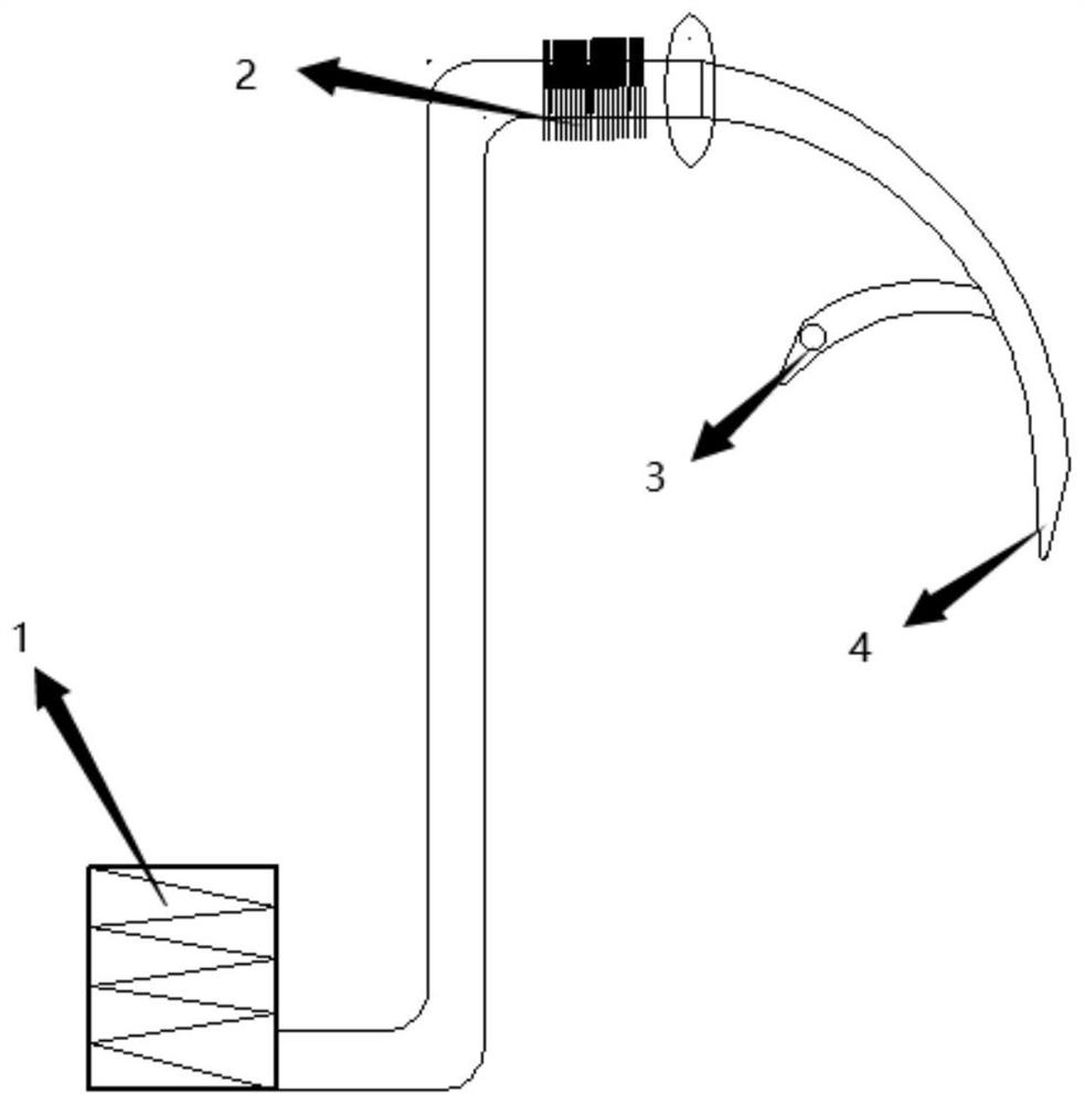
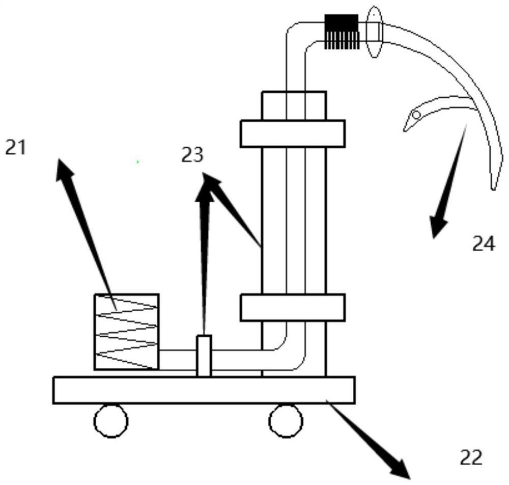
[0056] like figure 1 Shown, the bottom of the present invention is a compressible cylinder, and the connecting conduit is connected with the detection intubation again, and the detection intubation includes 3 detectors (cameras) and 4 intubation conduits.

[0057] When inserting the esophageal intubation, use this device to insert the male connector of ...


PUM


Abstract

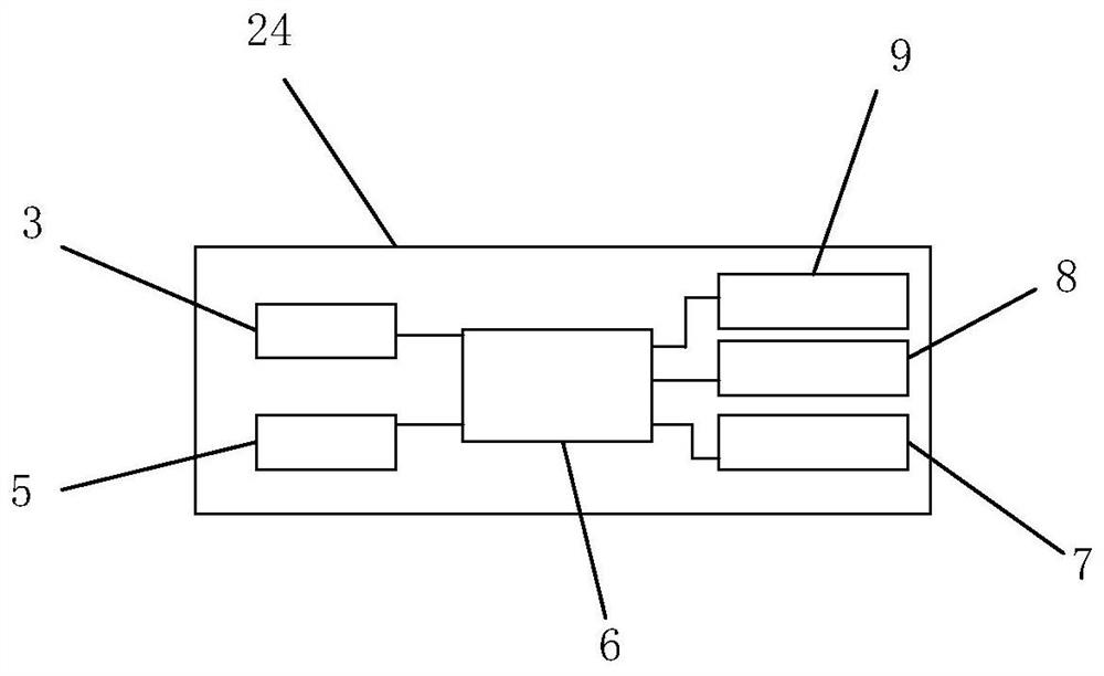
The invention belongs to the field of medical equipment, and discloses a digestive department esophageal intubation detection system and an image enhancement method, the digestive department esophageal intubation detection system comprises a camera, a master controller, an image enhancement module, a wireless communication module and an image cache module; the camera rotates in all directions and is used for full-angle camera shooting; the master controller is connected with the camera, the illuminating lamp, the image caching module, the image enhancement module and the wireless communication module through circuit lines. The image caching module is connected with the main controller and is used for caching the images in the esophagus acquired by the camera; the image enhancement module is used for enhancing the image in the esophagus acquired by the camera; and the wireless communication module is used for performing wireless transmission operation on the image in the esophagus acquired by the camera. The angle of the camera can be adjusted more effectively, wide imaging is achieved, a detection tube is omitted, multi-angle image information of an esophageal patient can be acquired, and discomfort of the patient is reduced.

Description

technical field [0001] The invention belongs to medical equipment, in particular to a detection system and an image enhancement method for an esophageal intubation tube in the department of gastroenterology. Background technique [0002] The current application of probing techniques in the esophagus is based on the anatomical differences between the esophagus and trachea. When inserting a catheter into a patient, it is often necessary to detect whether the position of the endotracheal tube is correct, and whether there will be an error into the esophagus. At this time, an endoesophageal intubation detection device is required for detection. There are now two detection devices for intubation in the esophagus. [0003] Through the above analysis, the problems and defects in the prior art are: [0004] One is a syringe-type esophageal detection device. There is an adapter in front of the syringe that can be connected to the endotracheal catheter. When the injection is connect...

Claims


Application Information

Patent Timeline
IPC IPC(8): G06T5/00G06T7/136H04N5/225H04N5/232H04N7/18A61B5/06A61B1/273A61B1/04
CPCG06T7/136H04N7/18A61B5/061A61B1/2733A61B1/04A61B1/00009A61B1/00016G06T2207/10016G06T2207/20021G06T2207/10068G06T2207/30021H04N23/00H04N23/555H04N23/67G06T5/92
Inventor 刘广明饶敏秦俊杰
Owner JILIN UNIV FIRST HOSPITAL
Learn More