
Facilitating transitioning between viewing native 2d and reconstructed 3D medical images

A medical image and medical imaging technology, applied in the field of medical imaging, can solve problems such as functional fusion

Pending Publication Date: 2019-12-06
GENERAL ELECTRIC CO
View PDF7 Cites 7 Cited by
  • Summary
  • Abstract
  • Description
  • Claims
  • Application Information

AI Technical Summary

Problems solved by technology

The current problem is the fusion of functions between these two types of independent applications

Method used


Image

  • Facilitating transitioning between viewing native 2d and reconstructed 3D medical images
  • Facilitating transitioning between viewing native 2d and reconstructed 3D medical images
  • Facilitating transitioning between viewing native 2d and reconstructed 3D medical images

Examples


Embodiment Construction

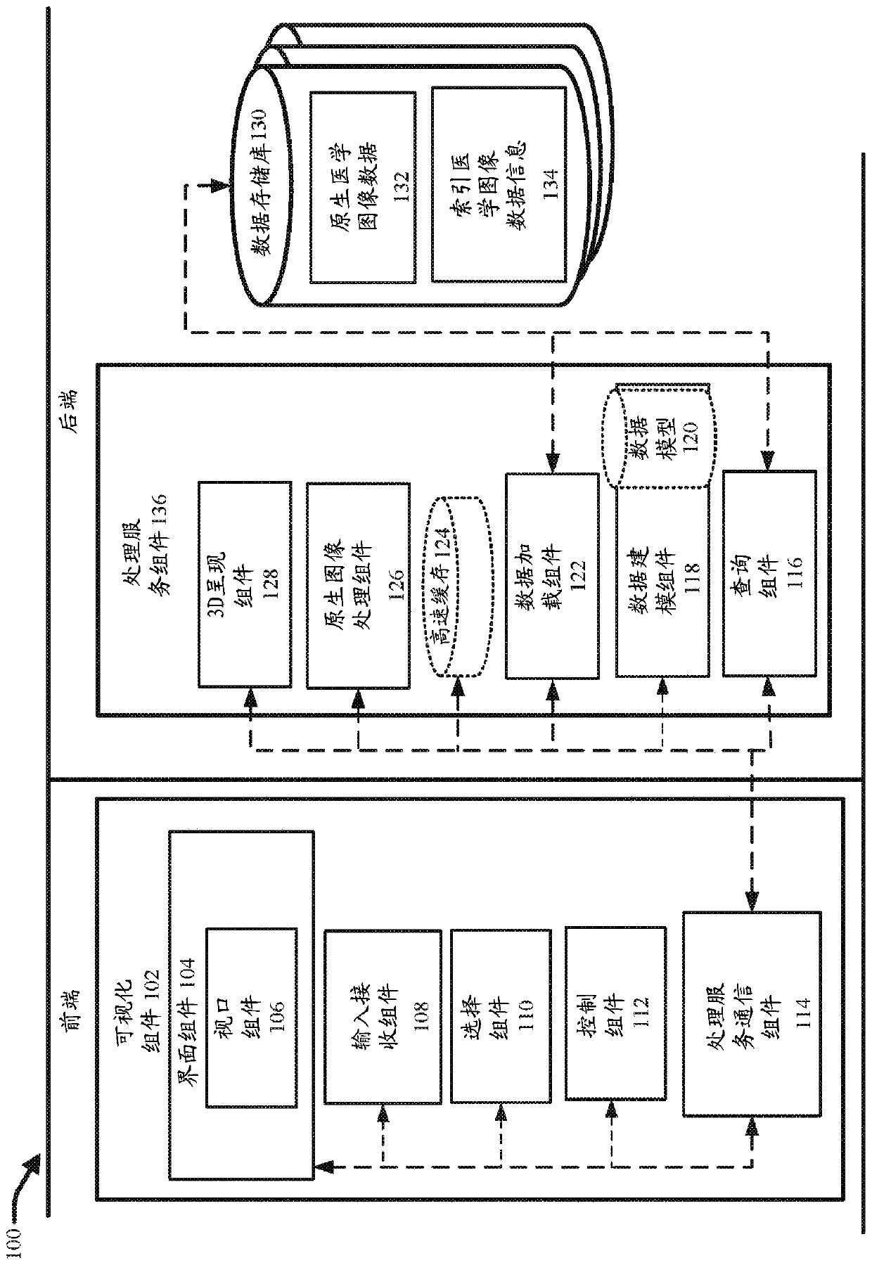
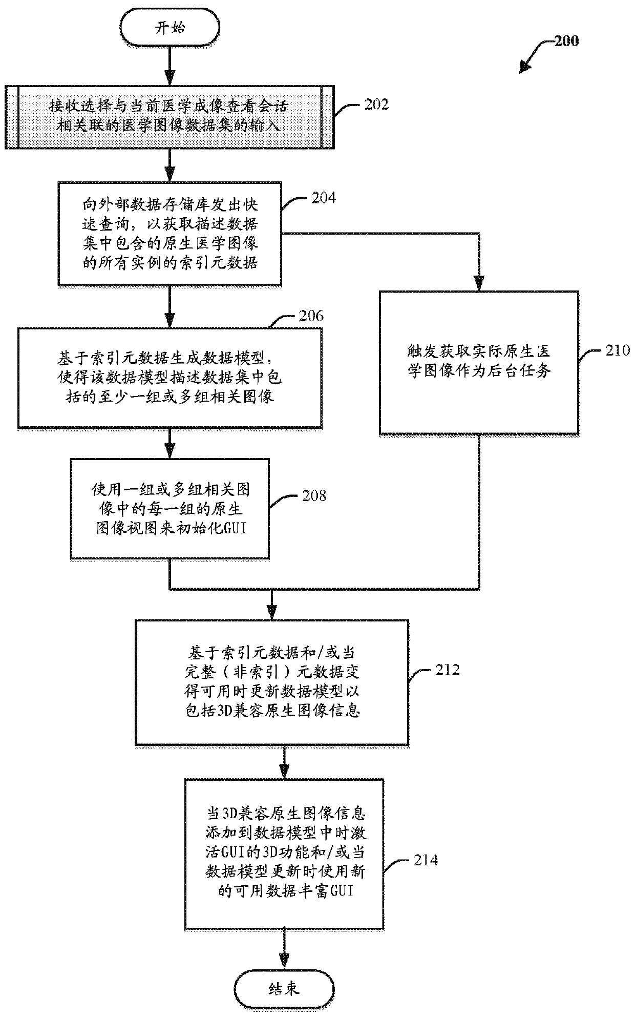
[0032]By way of introduction, the subject disclosure relates to systems, methods, apparatus, and computer-readable media that provide a medical imaging visualization application for viewing native and reconstructed medical images in the same display area or viewport of a GUI transition seamlessly. According to various embodiments, the medical imaging visualization application behaves as a native image viewer by default. In this regard, the visualization application can be configured to load native medical images associated with various types of imaging studies in any modality (e.g., x-ray, computed tomography (CT), mammography Radiography (MG), Magnetic Resonance Imaging (MRI), etc.), and display these native medical images in the GUI immediately as they come out of the acquisition system. For example, in one or more embodiments, after selecting a particular data set for viewing (e.g., a particular imaging study or group of imaging studies for a particular patient), the visua...


PUM


Abstract

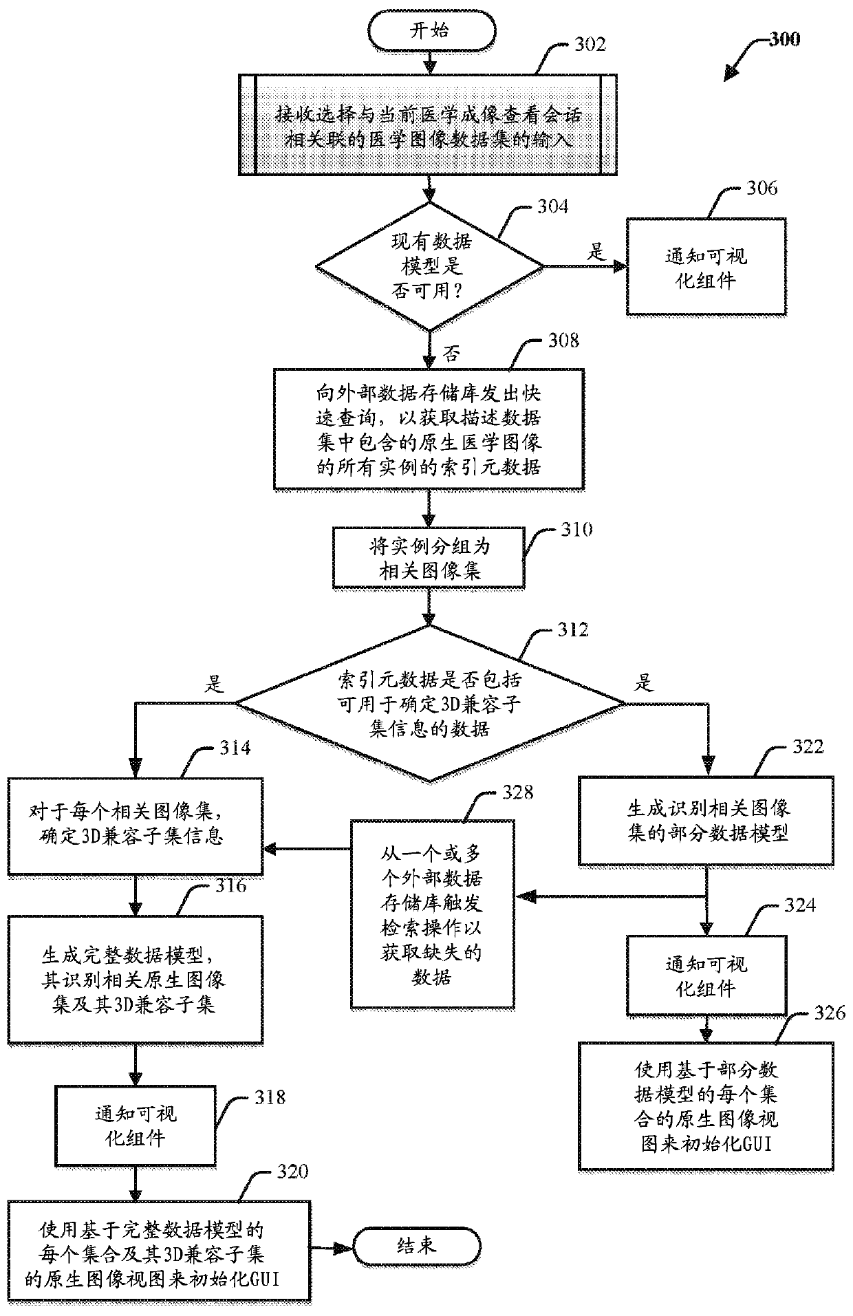
Medical imaging systems are provided that facilitate transitioning between viewing native and reconstructed medical images in the same viewport. In one embodiment, a method comprises retrieving indexed image metadata describing native medical images associated with a medical imaging data set selected for viewing. The method further comprises grouping the native medical images into one or more setsof related images, and for each set of the one or more sets of related images, sorting the related images into one or more subsets of 3D compatible images that can respectively be used to generate one or more 3D representations. The method further comprises generating a data model identifying the one or more sets of related images and the one or more subsets of 3D compatible images, and employingthe data model to facilitate transitioning between rendering the native medical images and the one or more 3D representations in the same viewport.

Description

technical field [0001] The present application relates generally to medical imaging, and more particularly to a medical imaging visualization system that facilitates transition between viewing native two-dimensional (2D) medical images and reconstructed three-dimensional (3D) medical images. Background technique [0002] Over the past decade, standard care has changed as radiologists have adopted reconstructed 3D medical image reading in their daily practice. These reconstructed medical images aid in the assessment and description of complex anatomy, injury and pathology. However, native 2D medical images that have been captured from medical imaging devices are still necessary as their interpretation tends to be faster. They are also the primary data that clinicians will examine to ensure that what they are looking at comes directly from the acquisition system, avoiding partial volume or inaccurate reconstruction artifacts. [0003] There are many medical viewer applicatio...

Claims


Application Information

Patent Timeline
IPC IPC(8): G16H30/40G16H40/60
CPCG06T19/00G06F16/51G06T2200/24G06T2210/41G06F3/04842G06T17/10G06T11/206G06F16/00
Inventor 尼古拉斯·德斯沃特科里内·英戈尔德佛科尼扎尔·奥德赫安托万·阿利奥蒂帕特里斯·赫沃让·拉巴尔
Owner GENERAL ELECTRIC CO
Learn More