Urine microalbumin detection method based on DNA aptamer and kit thereof
A technology for urinary microalbumin and nucleic acid aptamers, which is applied in the biological field and achieves the effects of wide application, high affinity and specificity, and simple and fast operation.
- Summary
- Abstract
- Description
- Claims
- Application Information
AI Technical Summary
Problems solved by technology
Method used
Image
Examples
Embodiment 1
[0032] Embodiment 1. according to the method of the present invention, adopt longer HSA nucleic acid aptamer (table 1, Cy3-HSA-apt) to the determination of the working curve that different concentrations of HAS are detected
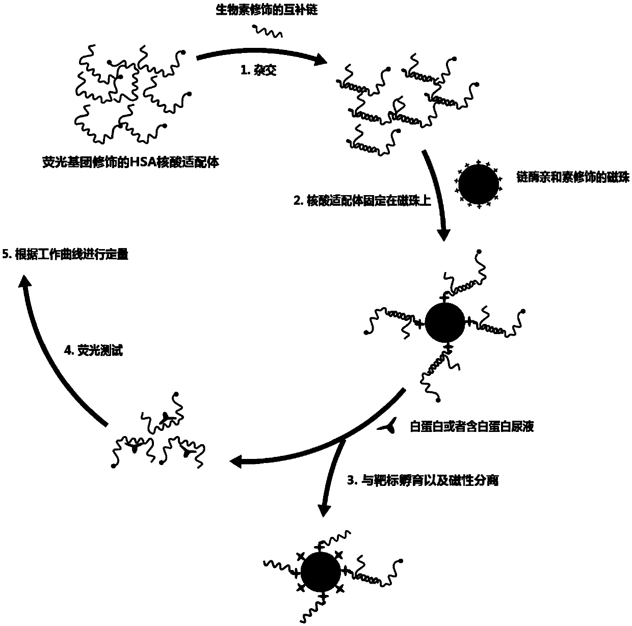
[0033] Specific methods include such as figure 1 The steps shown are as follows:
[0034] (1) Hybridization of nucleic acid aptamers and complementary probes: Cy3-HSA-apt (final concentration is 0.1 micromoles per liter (μM) and biotin-EG18-c-pool (Table 1, final concentration is 0.15 μM) Place in 1-fold concentrated binding and washing buffer solution (1×B&W) (5 millimolar per liter (mM) trishydroxymethylaminomethane (Tris), 0.5 mM ethylenediaminetetraacetic acid (EDTA), 1 mole per liter liter (M) sodium chloride (NaCl)), hybridization at a material ratio of 1:1.5: 95°C for 10 minutes, slowly cooled to room temperature.
[0035] (2) Immobilization of the nucleic acid aptamer on the magnetic beads: the glass bottle containing the streptavidin-coated mag...
Embodiment 2
[0040] Example 2. According to the method of the present invention, using a longer HSA nucleic acid aptamer (Table 1, Cy3-HSA-apt) to perform fluorescence detection on normal human urine with standard additions of 30nM and 300nM HSA respectively
[0041] (1) hybridization of nucleic acid aptamer and complementary probe: same as step (1) of embodiment 1;
[0042] (2) Immobilization of the nucleic acid aptamer on the magnetic beads: same as step (2) of Example 1;
[0043] (3) Divide the magnetic beads obtained in (2) into two groups: 1×Abb group: add HSA to the urine diluted 10 times (the final concentration of HSA is 0nM, 30nM, 300nM) to make the solution in each tube The final volume was 110 μL, mixed slowly, and incubated at room temperature for 1 hour.
[0044] (4) Fluorescence measurement: same as step (4) of Example 1.
[0045] The normal reference value range of HSA takes 24h urine albumin as the measurement standard, and its normal reference range is 30-300mg / 24h. The...
Embodiment 3
[0046] Embodiment 3. According to the method of the present invention, the determination of the working curve of detecting different concentrations of HSA using shorter HSA nucleic acid aptamers (Table 1, Cy3-HSA-apt-sh)
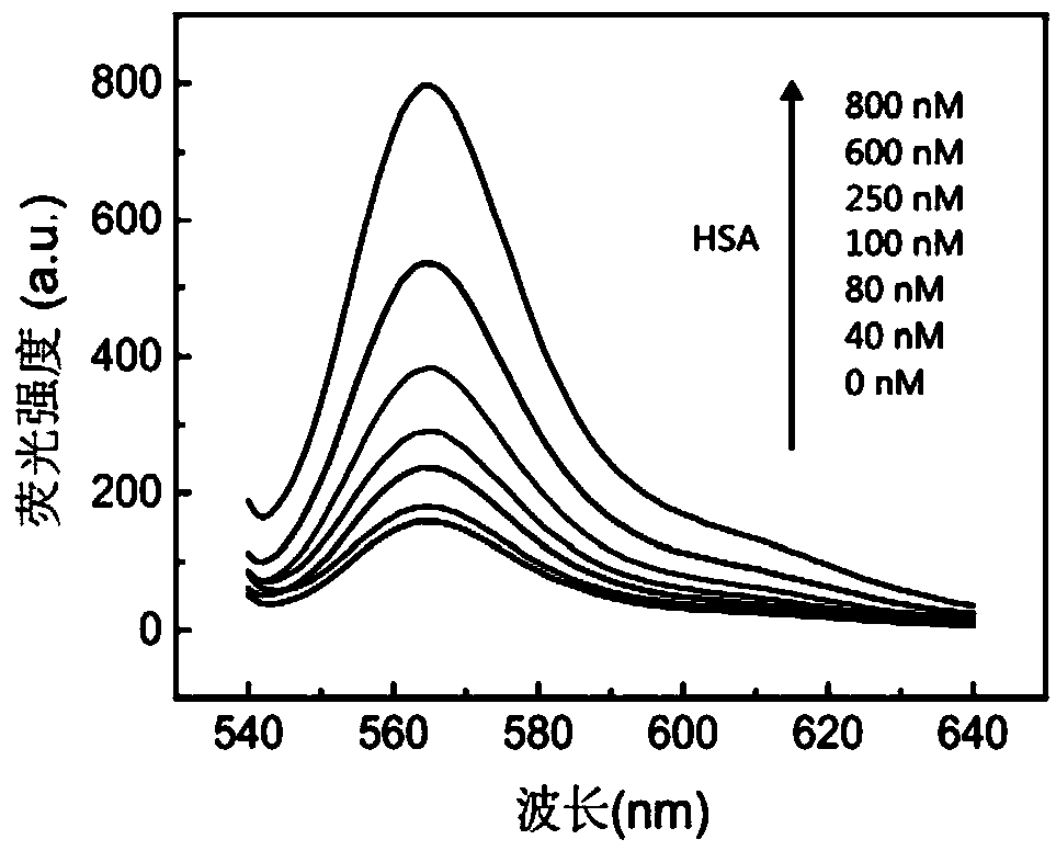
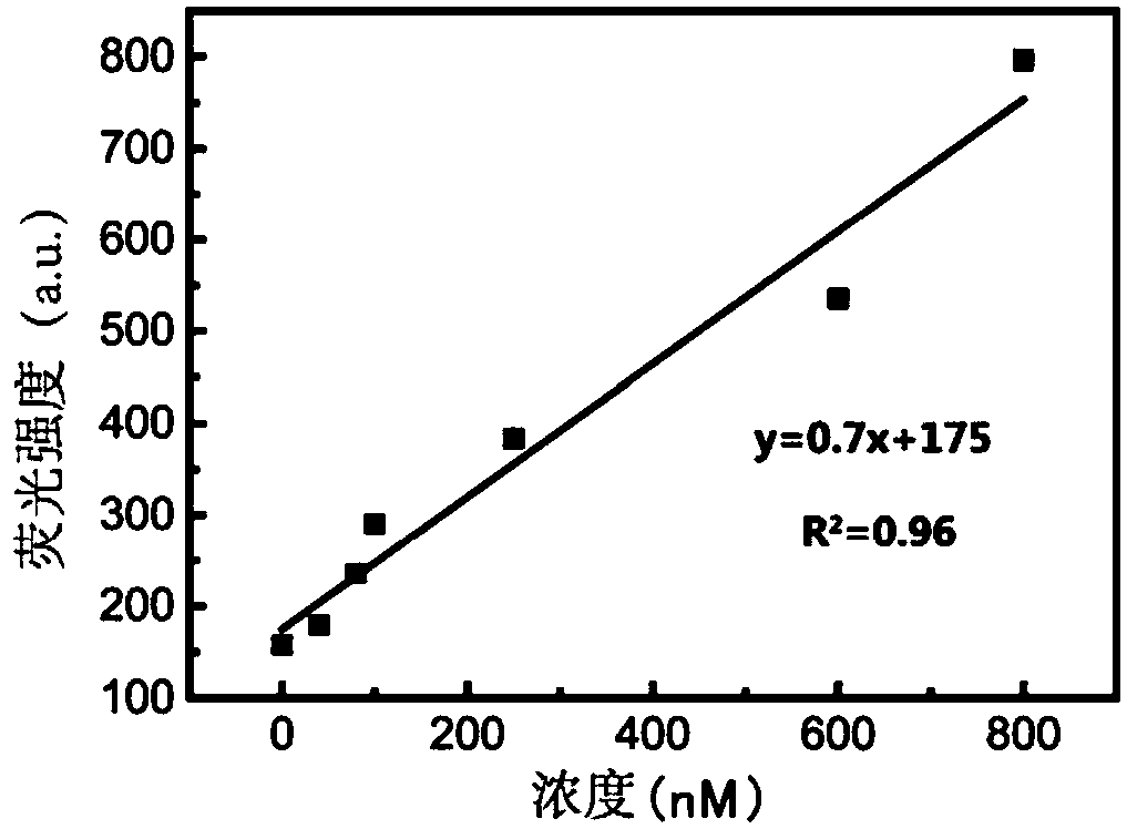
[0047] The experimental operation is the same as in Example 1, except that the HSA aptamer (Cy3-HSA-apt) used in Example 1 is replaced by a truncated HSA aptamer (Cy3-HSA-apt-sh), and The final concentration of HSA in the system is 0nM, 30nM, 80nM, 100nM, 300nM, 600nM, 800nM.
[0048] Such as Figures 4A-4B As shown, the fluorescence signal increases with the increase of HSA concentration, close to a linear relationship within 800nM HSA, and the detection limit is 80nM. The sensitivity of the detection was slightly lower than that of using a longer HSA aptamer (Cy3-HSA-apt).
PUM
 Abstract
Description
Claims
Application Information
 - R&D
- Intellectual Property
- Life Sciences
- Materials
- Tech Scout
- Unparalleled Data Quality
- Higher Quality Content
- 60% Fewer Hallucinations
Browse by: Latest US Patents, China's latest patents, Technical Efficacy Thesaurus, Application Domain, Technology Topic, Popular Technical Reports.
© 2025 PatSnap. All rights reserved.Legal|Privacy policy|Modern Slavery Act Transparency Statement|Sitemap|About US| Contact US: help@patsnap.com


