Culture method for umbilical cord blood CIK cells
A culture method and cord blood technology, which is applied in the field of cord blood CIK cell culture, can solve the problems of low proliferation rate and killing activity of CIK lymphocytes, complicated operation, etc., and achieve the promotion of adhesion and proliferation, high expansion, and avoid rejection The effect of the reaction
- Summary
- Abstract
- Description
- Claims
- Application Information
AI Technical Summary
Problems solved by technology
Method used
Image


Examples
Embodiment 1
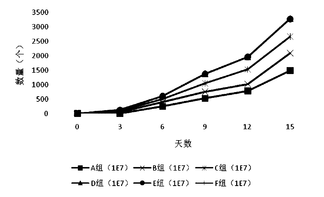
[0045] The method of this embodiment is as follows: extract umbilical cord blood mononuclear cells (CBMC) with lymphocyte separation medium, count the cells and divide them into six groups on average, including groups A, B, C, D, E, and F. Group A was cultured by the traditional method without RN, group B was cultured with RN (final concentration: 6.25 μg / mL), group C was cultured with RN (final concentration: 9.25 μg / mL), group D was cultured with RN ( The final concentration was 12.5 μg / mL) for coating culture, group E was used for coating culture (final concentration was 15.5 μg / mL), group F was cultured for coating with RN (final concentration was 18.5 μg / mL), trypan blue was used every 3 days The cell viability of each group was detected by staining, and the absolute number of cells was recorded. On the 7th, 14th, and 21st days of culture, the immunophenotypes of cells in each group were measured by flow cytometry. MTT assay was used to measure the killing rate of cells ...
Embodiment 2
[0111] This example is carried out with reference to step 6 of Example 1, the difference is that the final concentration of IFN-γ in the culture system is 500 IU / mL, and the final concentrations of IL-2 and IL-1a in the culture system are both 500 IU / mL, The final concentration of anti-CD3 monoclonal antibody in the culture system was 30ng / mL.
[0112] Table 5 Two groups of cell phenotypes under different culture time
[0113] CD3+CD56+ double positive D0 D3 D6 D9 D12 D15 H1 group 1% 5.8% 9.1% 27.3% 49.8% 66.4% H2 group 1% 6% 11.5% 30.5% 57.3% 75.3%
[0114] It can be seen from Table 5 that the final concentration of IFN-γ, IL-2 and IL-1α in the culture system is 500IU / mL, and when the final concentration of anti-CD3 monoclonal antibody in the culture system is 30ng / mL, the The final concentration of γ, IL-2, and IL-1α in the culture system was 1000IU / mL, and the final concentration of anti-CD3 monoclonal antibody in the culture syste...
Embodiment 3
[0119] This example is carried out with reference to Example 1, the difference is that the final concentration of IFN-γ in the culture system is 1500IU / mL, the final concentrations of IL-2 and IL-1a in the culture system are both 1500IU / mL, anti-CD3 monoclonal The final concentration of the cloned antibody in the culture system was 70ng / mL.
[0120] Table 7 Two groups of cell phenotypes under different culture time
[0121] CD3+CD56+ double positive D0 D3 D6 D9 D12 D15 H1 group 1% 6.2% 9.4% 26.9% 50.9% 68.8% H2 group 1% 6.3% 11.5% 32% 59.1% 76.9%
[0122] It can be seen from Table 7 that the final concentration of IFN-γ, IL-2 and IL-1α in the culture system is 1500IU / mL, and when the final concentration of anti-CD3 monoclonal antibody in the culture system is 70ng / mL, it can react with IFN-γ The final concentration of IL-2 and IL-1α in the culture system was 1000IU / mL, and the final concentration of anti-CD3 monoclonal antibody in the...
PUM
 Abstract
Description
Claims
Application Information
 - R&D Engineer
- R&D Manager
- IP Professional
- Industry Leading Data Capabilities
- Powerful AI technology
- Patent DNA Extraction
Browse by: Latest US Patents, China's latest patents, Technical Efficacy Thesaurus, Application Domain, Technology Topic.
© 2024 PatSnap. All rights reserved.Legal|Privacy policy|Modern Slavery Act Transparency Statement|Sitemap