
Medical ultrasonic image segmentation method

An ultrasound image, medical technology, applied in the field of medical ultrasound image segmentation

Active Publication Date: 2020-05-19
UNIV OF ELECTRONICS SCI & TECH OF CHINA +1
View PDF15 Cites 2 Cited by
  • Summary
  • Abstract
  • Description
  • Claims
  • Application Information

AI Technical Summary

Problems solved by technology

However, traditional CNNs cannot reasonably propagate low-level features to high-level

Method used


Image

  • Medical ultrasonic image segmentation method
  • Medical ultrasonic image segmentation method
  • Medical ultrasonic image segmentation method

Examples


Embodiment Construction

[0024] Below in conjunction with accompanying drawing and emulation the present invention is described in detail:

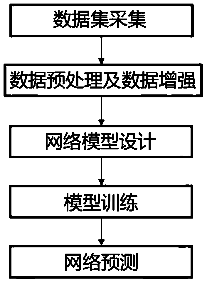
[0025] The present invention provides a network segmentation method based on ultrasound images of thyroid nodules, which includes 5 steps, mainly including 5 modules of data set acquisition, image preprocessing, network model construction, network training, network testing and evaluation, and its flow chart is as follows Figure 1 shows. In this embodiment, the specific steps are as follows:

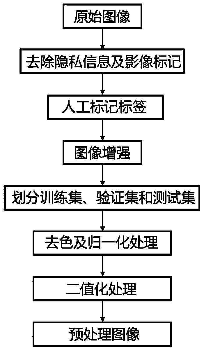
[0026] 1. Preprocess the ultrasonic image data to be segmented to obtain training set data and test set data. The data processing flow is shown in Figure 2.

[0027] 1) Remove private information and medical imaging equipment marks, and filter out the original ultrasound images that have not been manually marked by radiologists;

[0028] 2) Manually mark labels under the guidance of ultrasound imaging physicians;

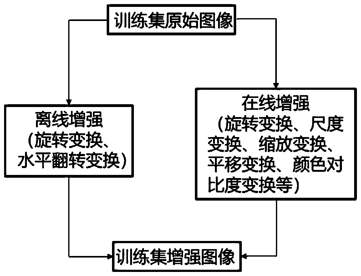
[0029] 3) Enhance the image quality under the premise of ...


PUM


Abstract

The invention belongs to the technical field of deep learning computer vision and medical information processing, and particularly relates to a medical ultrasonic image segmentation method. Accordingto the method, on the basis of a general image segmentation neural network model, multiple novel technologies such as a multiple-input-multiple-output technology, a hole convolution technology and small sample medical data enhancement are fused, the problems of difficult pain points such as small sample learning, low ultrasonic image contrast and nodule edge blurring are mainly solved, and an optimal segmentation strategy of the method is obtained.

Description

technical field [0001] The invention belongs to the technical field of deep learning computer vision and medical information processing, and in particular relates to a medical ultrasound image segmentation method. Background technique [0002] With the advancement of science and technology, medical imaging technology has made great progress. Ultrasound imaging technology is of great value in preventive diagnosis and treatment due to its advantages of simple operation, no radiation damage, and low cost. At present, the segmentation of regions of interest in medical images is the basis for image analysis and lesion identification. Manual segmentation is widely used clinically to segment ultrasound images, and experienced clinicians manually outline areas of interest based on their professional knowledge. However, manual segmentation is not only time-consuming and extremely dependent on the professional skills and rich experience of doctors, but also due to the blurred edges a...

Claims


Application Information

Patent Timeline
IPC IPC(8): G06T7/10G06T5/00
CPCG06T7/10G06T2207/10132G06T2207/20081G06T2207/20084G06T2207/20221G06T5/90Y02T10/40
Inventor 车博袁浩瀚罗亮陈智方俊熊雯
Owner UNIV OF ELECTRONICS SCI & TECH OF CHINA
Learn More