Method for detection and display of extravasation and infiltration of fluids and substances in subdermal or intradermal tissue

a technology of fluids and substances, applied in the field of medical devices and procedures, can solve the problems of tissue damage, extravasation and infiltration, discoloration of the skin,

Inactive Publication Date: 2006-08-03
MEDRAD INC.
View PDF24 Cites 47 Cited by
  • Summary
  • Abstract
  • Description
  • Claims
  • Application Information

AI Technical Summary

Problems solved by technology

Extravasation describes leaking of intravascular fluids or medicines that may, in a worst case scenario, cause tissue damage.
Incidents that cause extravasation and infiltration include improper venipuncture, such as a transfixation of a vein, rupture of the vasculature, perhaps due to weakened vascular walls in patients of advanced age, disease states, abrasion by the cannula, or the administration of a toxic agent.
Problems with extravasation and infiltration may include bruising, discoloration of the skin or discomfort to the patient, or more serious problems associated with thrombosis, bleeding leading to hypovolemia, and tissue toxicity, such as in the administration of toxic substances associated with chemotherapy wherein the concentration of a toxic substance is carefully monitored to ensure dilution of the substance to appropriate levels during administration.
Extravasation into the spaces surrounding a blood vessel poses very serious problems of local tissue toxicity and / or possible necrosis depending upon the agent, and a lack of accurate delivery of metered dosage into the patient's vascular system.
Treatment of suspected extravasation may be costly and time consuming.
First, an effort must be made to determine whether an extravasation or infiltration has occurred, the material injected, the injection location, and the amount of injected volume into the tissue.
These prior methods as a whole exhibit significant limitations in that they cause direct obstructions at the site of injection and extravasation or infiltration thereby preventing visualization and palpation of the tissue, a critical step by the healthcare provider under all circumstances.
This method, along with the other prior art for detection of extravasation, only discloses a technique for generation of electronic signals for processing and interpretation of extravasation or infiltration events; they do not teach a method of immediate visual display of the actual physical features in the form of contrast images of the extravasating or infiltrating fluids.
These methods may indicate vein or artery location, but do not allow for the detection of extravasation or infiltration in direct visual respect to the vasculature.
However, the device and method taught in the '046 patent does not address the sensing, and display the presence, of extravasated or infiltrated fluids and substances into subdermal or intradermal tissues.

Method used


Image

  • Method for detection and display of extravasation and infiltration of fluids and substances in subdermal or intradermal tissue
  • Method for detection and display of extravasation and infiltration of fluids and substances in subdermal or intradermal tissue
  • Method for detection and display of extravasation and infiltration of fluids and substances in subdermal or intradermal tissue

Examples


Embodiment Construction

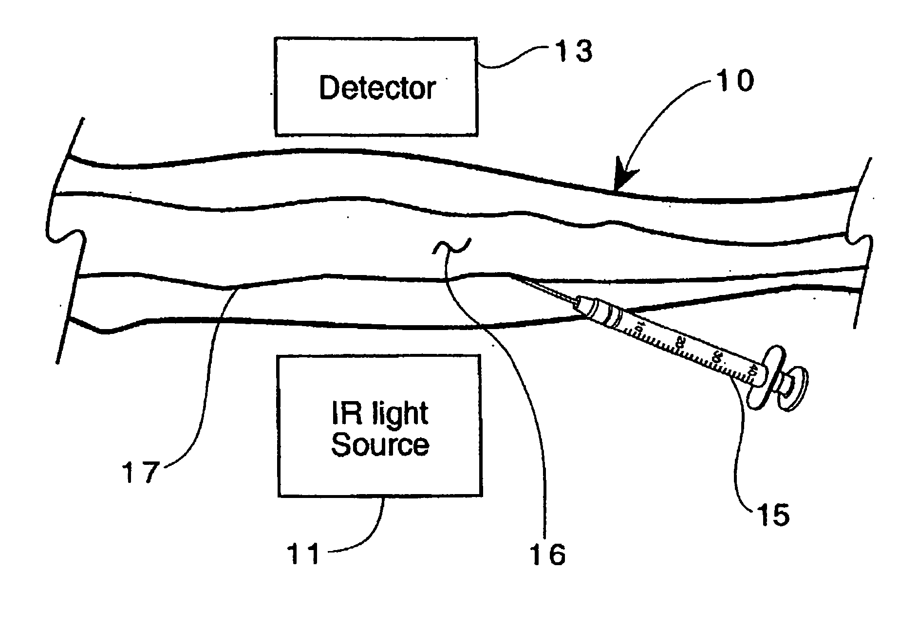
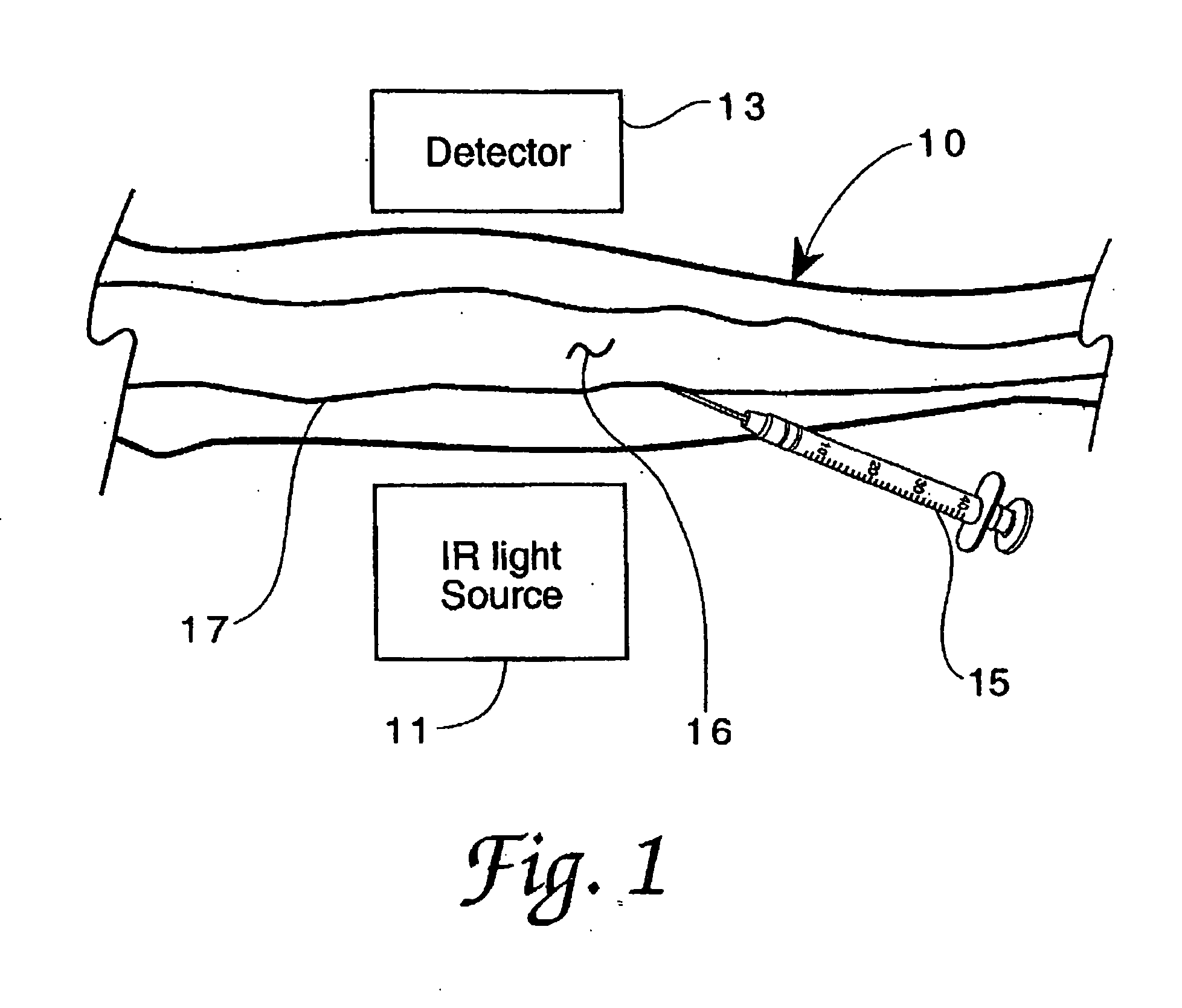
[0027] Referring now to the drawings, FIG. 1 illustrates the method of the invention in one of its embodiments for imaging extravasation or infiltration, wherein a surface area of the body, such as forearm 10, is placed near an NIR light source 11 and between source 11 and detector 13 (in the transillumination mode of the invention), in order to perform a particular procedure, such as in the administration of a specific substance into the vasculature (artery or vein 17) using IV instrument 15. Source 11 preferably emits light in the wavelength range of about 0.3 to 1.0 microns (μm), and detector 13 is sensitive to light in that range. Alternatively, source 11 could be placed to directly illuminate forearm 10 such that a reflected NIR image of the infusion area 16 at instrument 15 is viewed. Source 11 can also be placed near the area of interest of forearm 10 such that scattered light is used to illuminate the area of interest. Detector 13 is focused upon the area of interest and doe...


PUM


Abstract

A method for the real-time visualization and detection of extravasated and or infiltrated fluid and substances, including blood, that occur near the cannulation site of an injection is described wherein illumination or transillumination with near infrared light is used to image the contrast in real-time between absorbing and nonabsorbing subdermal and intradermal structures of blood vessels and remaining surrounding tissue, foreign substances and other structures in order to establish a baseline image of the body area of interest, and any new image is monitored and compared with the baseline image to detect the extravasation and / or infiltration of fluids and substances, including blood, around a vein or artery into the subdermal or intradermal tissue.

Description

FIELD OF THE INVENTION [0001] The invention relates generally to medical devices and procedures, and more particularly to procedures for the real-time detection and visualization of infiltrated or extravasated substances, including blood, that occur in subdermal or intradermal tissues near the cannulation site of an injection procedure, such as in the intravascular delivery or extraction of various substances or media. BRIEF DESCRIPTION OF RELATED ART [0002] The following information is provided to assist the reader to understand the invention disclosed below and at least some of the applications in which it will typically be used. In addition, any references set forth herein are intended merely to assist in such understanding. Inclusion of a reference herein, however, is not intended to constitute an admission that the reference is available as prior art with respect to the invention. [0003] During intravascular administration of substances, a portion of the substances may escape f...

Claims


Application Information

Patent Timeline
Patent Type & Authority Applications(United States)
IPC IPC(8): A61B6/00
CPCA61B5/0059
Inventor KALAFUT, JOHN F.GRIFFITHS, DAVID M.UBER, ARTHUR E. III
Owner MEDRAD INC.
Why Eureka
Try Eureka