Tumor component labeling method, device and equipment and storage medium

A tumor and labeling technology, applied in the field of image processing, can solve problems such as error-prone, cumbersome tumor components, and heavy workload, and achieve the effects of improving accuracy and efficiency, high precision, and saving labor costs

Pending Publication Date: 2020-05-19
上海镜观生物科技有限公司
View PDF8 Cites 1 Cited by
  • Summary
  • Abstract
  • Description
  • Claims
  • Application Information

AI Technical Summary

Problems solved by technology

[0003] Tumor components (including tumor cells, tumor microenvironment, and heterogeneous tumor cells, etc.) are marked in pathological sections mainly through special staining or sequencing. It is very cumbersome to identify the stained tumor components by naked eyes after scanning pathological sections into digital sections. Heavy workload and error-prone

Method used


Image

  • Tumor component labeling method, device and equipment and storage medium
  • Tumor component labeling method, device and equipment and storage medium
  • Tumor component labeling method, device and equipment and storage medium

Examples


Embodiment Construction

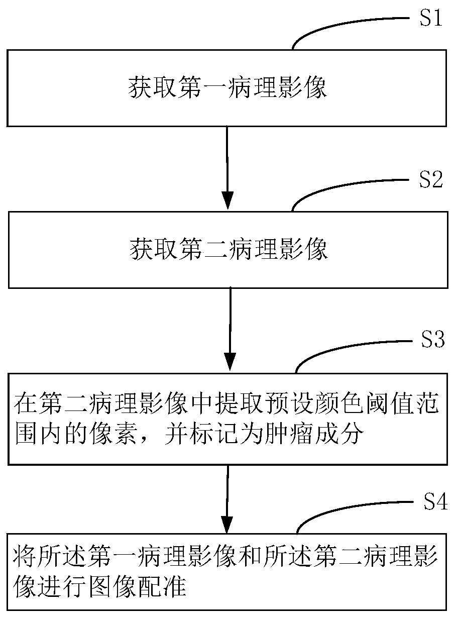
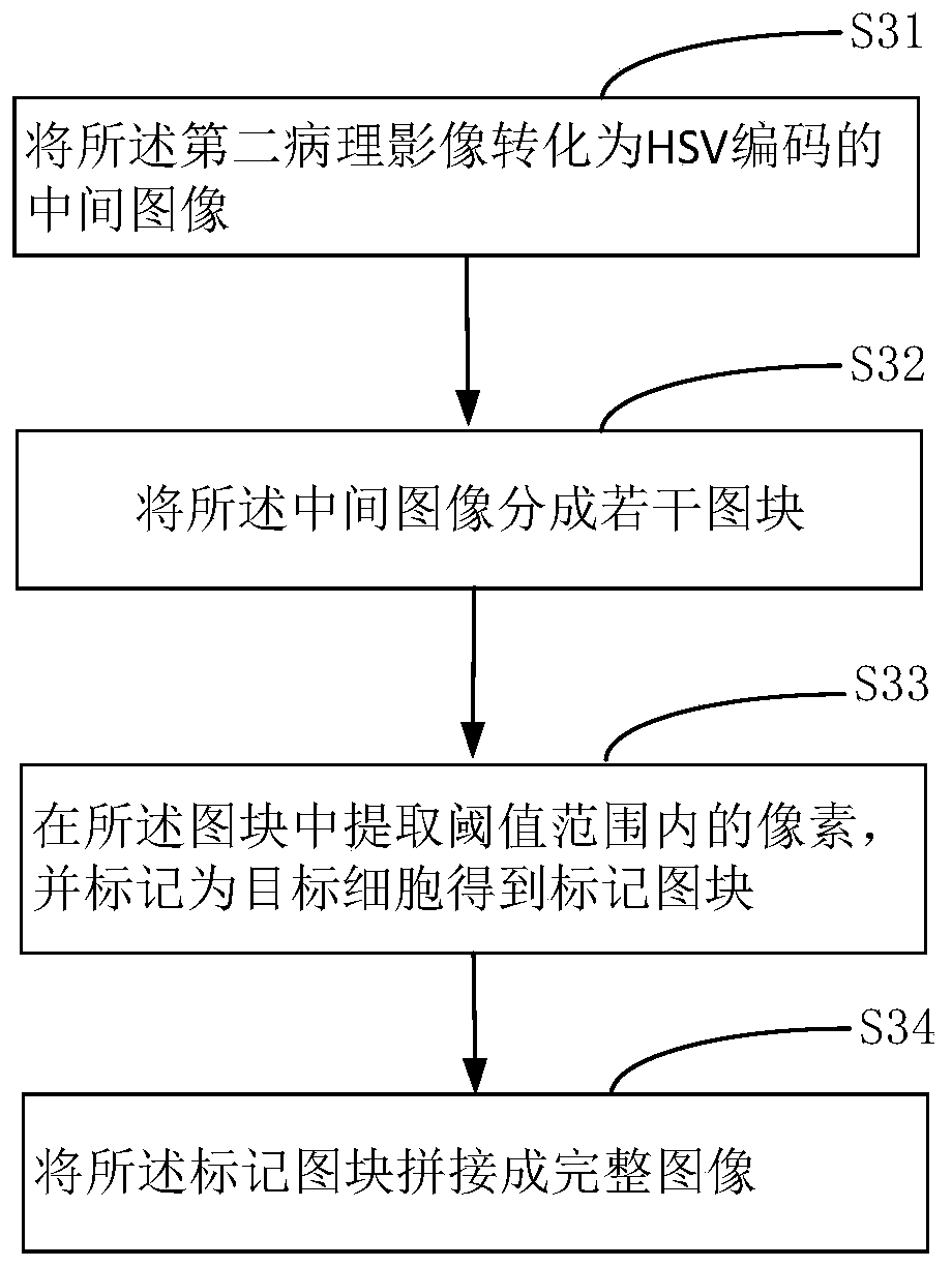
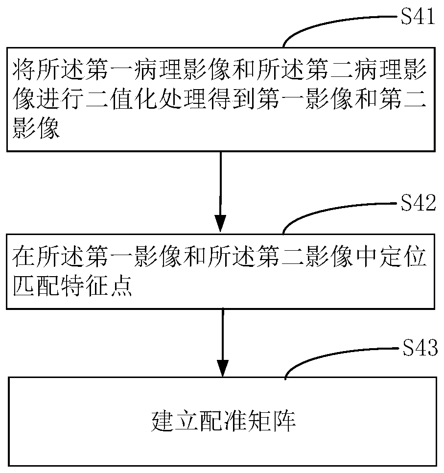
[0049] The conception and technical effects of the present invention will be clearly and completely described below in conjunction with the embodiments, so as to fully understand the purpose, features and effects of the present invention. Apparently, the described embodiments are only some of the embodiments of the present invention, rather than all of them. Based on the embodiments of the present invention, other embodiments obtained by those skilled in the art without creative efforts belong to The protection scope of the present invention.

[0050] In the description of the present invention, if it involves orientation description, for example, the orientation or positional relationship indicated by "upper", "lower", "front", "back", "left", "right" etc. is based on the The orientation or positional relationship is only for the convenience of describing the present invention and simplifying the description, but does not indicate or imply that the device or element referred ...


PUM


Abstract

The invention discloses a tumor component labeling method, device and equipment and a storage medium, and the method comprises the steps: obtaining a first pathological image, wherein the first pathological image is an image obtained through the digitalized scanning of a pathological section after HE staining; acquiring a second pathological image, wherein the second pathological image is an imageobtained by performing HE fading on the pathological section, then performing immunohistochemical staining and then performing digital scanning; extracting pixels in a preset color threshold range from the second pathological image, and marking the pixels as tumor components; and performing image registration on the first pathological image and the second pathological image. Tumor components canbe automatically marked in pathological images, manual marking is not needed, the marking speed is high, and the precision is high.

Description

technical field [0001] The present invention relates to the field of image processing, in particular to a tumor component labeling method, device, equipment and storage medium. Background technique [0002] Digital pathological slide is to quickly scan the entire slide with full information and all directions, turning the traditional materialized slide into a new generation of digital pathological slice. [0003] Tumor components (including tumor cells, tumor microenvironment, and heterogeneous tumor cells, etc.) are marked in pathological sections mainly through special staining or sequencing. It is very cumbersome to identify the stained tumor components by naked eyes after scanning pathological sections into digital sections. A lot of work and error-prone. Contents of the invention [0004] The present invention aims to solve at least one of the technical problems existing in the prior art. For this reason, the present invention proposes a method for labeling tumor co...

Claims


Application Information

Patent Timeline
IPC IPC(8): G16H30/20G16H30/40G06T7/33
CPCG16H30/20G16H30/40G06T7/33
Inventor 于观贞陈颖高云姝王进祥赵亮刘家琪吴湛
Owner 上海镜观生物科技有限公司
Why Eureka
Try Eureka