
Gland medical image display method, interaction method, and storage medium

A medical image and display method technology, applied in the fields of recognition, medical image processing, and display, can solve the problems that affect the ease of use of the diagnostic system, lack of multiple display forms, and multiple interactive modes

Active Publication Date: 2020-07-17
HANGZHOU YITU MEDIAL TECH CO LTD
View PDF9 Cites 10 Cited by
  • Summary
  • Abstract
  • Description
  • Claims
  • Application Information

AI Technical Summary

Problems solved by technology

In addition, in the products that support single lesion corresponding to multiple signs, there is currently a lack of multiple display forms and multiple interactive modes, which affects the ease of use of the diagnostic system

Method used


Image

  • Gland medical image display method, interaction method, and storage medium
  • Gland medical image display method, interaction method, and storage medium
  • Gland medical image display method, interaction method, and storage medium

Examples


Embodiment Construction

[0052] In order to make the purpose, technical solutions and advantages of the embodiments of the present disclosure clearer, the technical solutions of the embodiments of the present disclosure will be clearly and completely described below in conjunction with the drawings of the embodiments of the present disclosure. Apparently, the described embodiments are some of the embodiments of the present disclosure, not all of them. Based on the described embodiments of the present disclosure, all other embodiments obtained by persons of ordinary skill in the art without creative effort fall within the protection scope of the present disclosure.

[0053] Unless otherwise defined, the technical terms or scientific terms used in the present disclosure shall have the usual meanings understood by those skilled in the art to which the present disclosure belongs. "Comprising" or "comprising" and similar words mean that the elements or items appearing before the word include the elements o...


PUM


Abstract

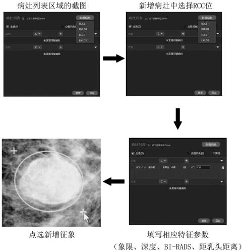
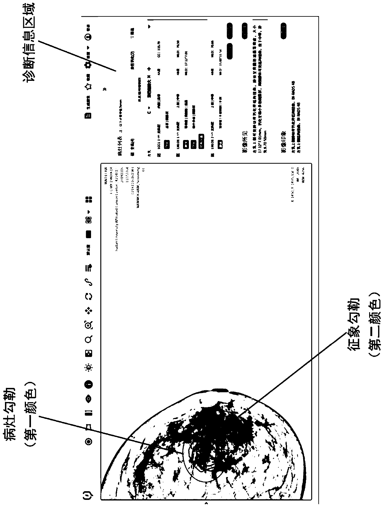
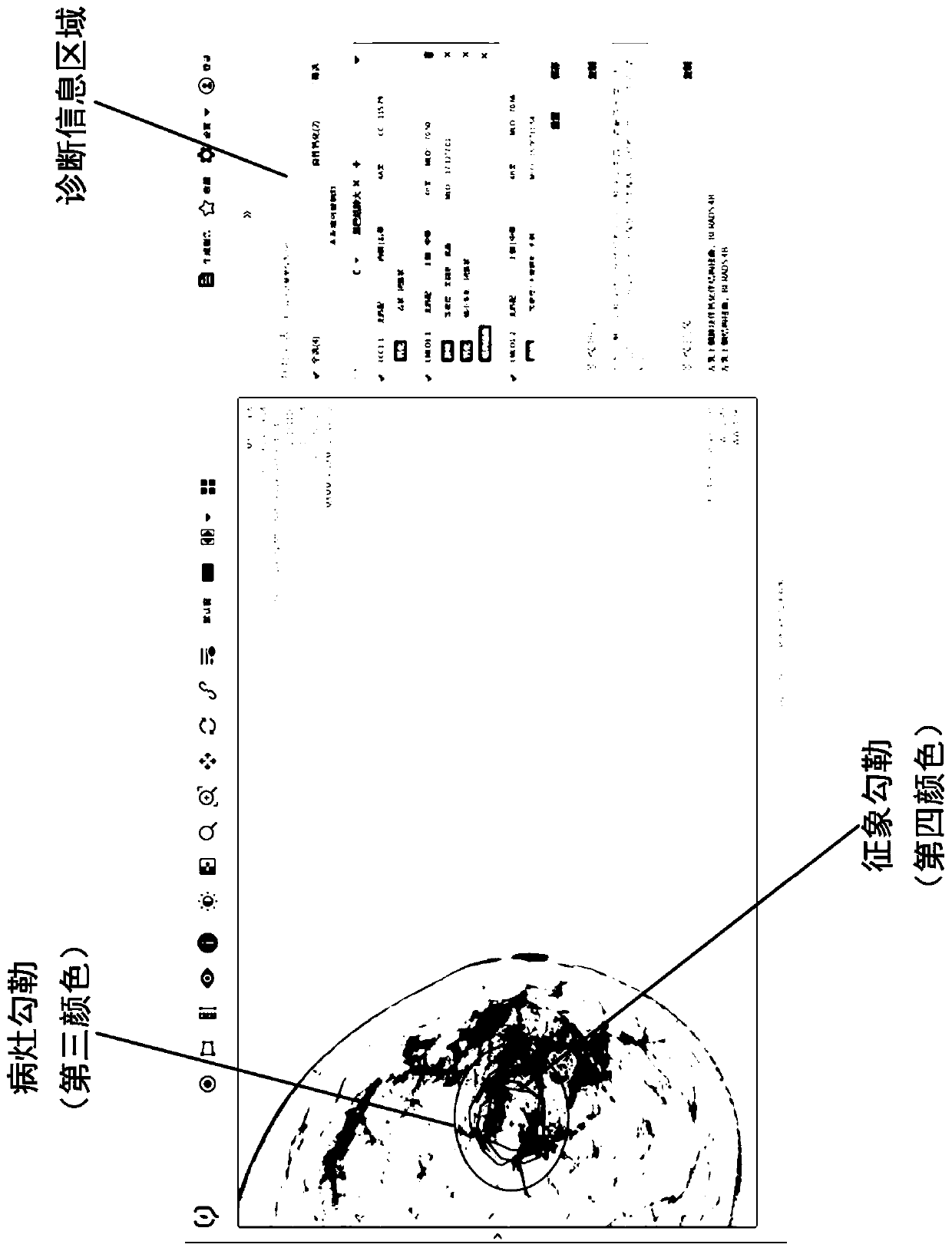
The disclosure relates to a gland medical image display method, a gland medical image diagnosis interface interaction method, and a computer-readable storage medium. The display method includes: defining an infection focus area and representing a gland medical image containing the infection focus area in response to the input operation of infection focus feature parameters of the gland; in response to the selection operation of an operation body on the gland medical image, representing the infection focus and symptoms with different markers. The interaction method comprises: in response of theinput operation of the feature parameters, providing different interaction modes on the gland medical image containing the infection focus area; in response of the selection operation, representing the infection focus and symptoms with different markers. According to the technical scheme, under the application status of single infection focus corresponding to multiple symptoms, an infection focuslist includes the infection focus items and symptoms items, which are corresponded to the infection focus markers and symptom markers inside the infection focus in the image in a one-to-one manner, thereby greatly maintaining the usability of the system.

Description

technical field [0001] The present disclosure relates to the technical fields of medical image processing, identification, and display, and in particular to a display method of glandular medical images, an interactive method of glandular medical image diagnosis interface, and a computer-readable storage medium. Background technique [0002] In the prior art, the diagnostic analysis for lesions and signs only supports single lesions corresponding to single signs, that is, there is only a single sign in a single lesion marker in a medical image of the gland. In the lesion list, there is only one sign type and corresponding physiological parameters in a single lesion. In addition, among the products that support single lesion corresponding to multiple signs, there is currently a lack of multiple display forms and multiple interactive modes, which affects the ease of use of the diagnostic system. Contents of the invention [0003] The present disclosure intends to provide a d...

Claims


Application Information

Patent Timeline
IPC IPC(8): G16H30/40G16H15/00G06F3/0484
CPCG16H30/40G16H15/00G06F3/04845G06F3/04842G06F3/04847
Inventor 石磊倪波
Owner HANGZHOU YITU MEDIAL TECH CO LTD
Learn More