MRI tumor optimal segmentation method and system based on multi-modal image fusion

A multi-modal image and image fusion technology, applied in image analysis, image enhancement, image data processing and other directions, can solve the problems of low segmentation efficiency and rough segmentation results, and achieve the effect of good feature representation and good performance.

Active Publication Date: 2020-09-01
复影(上海)医疗科技有限公司
View PDF4 Cites 10 Cited by
  • Summary
  • Abstract
  • Description
  • Claims
  • Application Information

AI Technical Summary

Problems solved by technology

[0007] For the existing traditional segmentation method, it is necessary to understand the image through human subjective consciousness, so as to extract specific feature information, such as gray level information, texture information and symmetry information, to realize the segmentation of brain tumors. The result can only be better for specific images. Segmentation results, and the technical problems that the segmentation results are too rough and the segmentation efficiency is low, the present invention provides an MRI tumor optimal segmentation network based on multimodal image fusion

Method used


Image

  • MRI tumor optimal segmentation method and system based on multi-modal image fusion
  • MRI tumor optimal segmentation method and system based on multi-modal image fusion
  • MRI tumor optimal segmentation method and system based on multi-modal image fusion

Examples


Embodiment Construction

[0043] The present invention will be described in detail below in conjunction with specific embodiments. The following examples will help those skilled in the art to further understand the present invention, but do not limit the present invention in any form. It should be noted that those skilled in the art can make several changes and improvements without departing from the concept of the present invention. These all belong to the protection scope of the present invention.

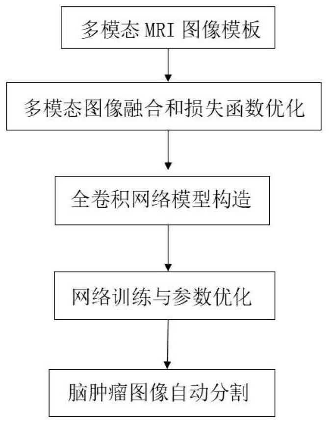
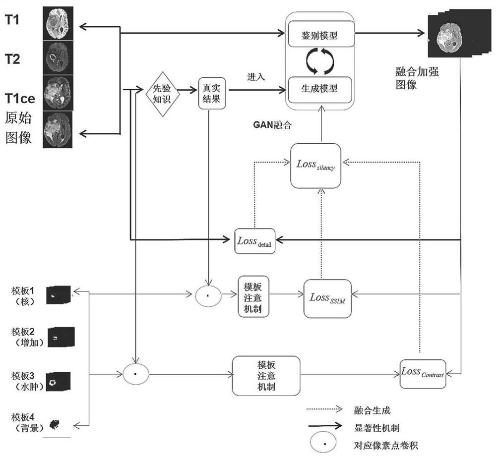
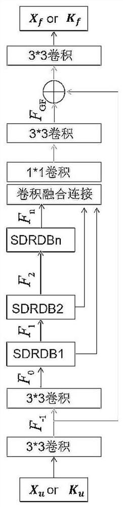
[0044]The invention combines medical images and deep learning algorithms to construct the generation of multimodal MRI brain tumor images and complete the segmentation of brain tumor MRI images. This MRI tumor optimal segmentation based on multimodal image fusion will have an important impact in the field of medical imaging. The present invention proposes an improved network structure based on U-Net, which integrates SE-Net network, and adaptively recalibrates the characteristic response of the channel ...


PUM


Abstract

The invention provides an MRI tumor optimal segmentation method and system based on multi-modal image fusion. The MRI tumor optimal segmentation method comprises the steps that step 1, creating an MRItumor multi-modal image fusion network step 2, constructing a multi-modal 3D network for enhancing tumor image segmentation; step 3, constructing a significance loss function based on GAN image fusion; step 4, constructing a Mask attention mechanism contrast loss function; step 5, constructing an SSIM loss function; and step 6, performing MRI tumor optimal segmentation according to the MRI tumormulti-modal image fusion network, the multi-modal 3D network and the three loss functions. When a deep architecture is trained, one remaining unit helps. Feature accumulation is carried out by using arecursive residual convolution layer, so that better feature representation is provided for a segmentation task; a U-NET system structure with the same network parameters and good performance is designed for medical image segmentation.

Description

technical field [0001] The present invention relates to the technical field of medical image segmentation, in particular to an MRI tumor optimal segmentation method and system based on multimodal image fusion. In particular, it relates to a 3D U-Net brain tumor segmentation method based on multimodal medical image fusion and loss function optimization. Background technique [0002] Multimodal MRI (nuclear magnetic resonance) images are an important diagnostic tool for the assessment and treatment of brain tumors. At present, compared with natural images, the resolution of medical imaging is relatively low, and the cost of medical imaging is relatively high, and the amount of data is relatively small . The threshold for labeling medical images is high, and only experienced and senior physicians can accurately label 3D images. Therefore, data samples and labels that can be used for computer science research are very scarce. The lack of data is one of the difficulties in the ...

Claims


Application Information

Patent Timeline
Patent Type & Authority Applications(China)
IPC IPC(8): G06T7/00G06T7/11G06T7/90G06N3/04G06K9/62G06K9/32
CPCG06T7/0012G06T7/11G06T7/90G06T2207/20081G06T2207/20084G06T2207/10088G06T2207/30016G06T2207/30096G06V10/25G06V2201/032G06N3/045G06F18/22G06F18/25Y02T10/40
Inventor 耿道颖于泽宽李郁欣尹波张军吴昊耿岩胡斌杨丽琴
Owner 复影(上海)医疗科技有限公司
Why Eureka
Try Eureka