
ELISPOT detection kit for detecting brucellosis and application of ELISPOT detection kit
A detection kit and technology for brucellosis, applied in the direction of measuring devices, instruments, scientific instruments, etc., can solve the problem of inability to detect brucella markers, detect brucella infection, and interfere with the accuracy of results To achieve the effect of truly reflecting the immune level in the body, objective test results, and improving specificity and sensitivity
- Summary
- Abstract
- Description
- Claims
- Application Information
AI Technical Summary
Problems solved by technology
Method used
Image

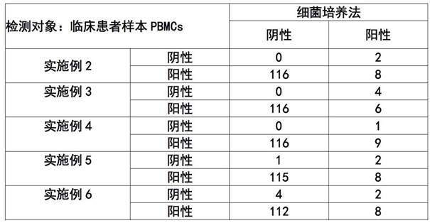
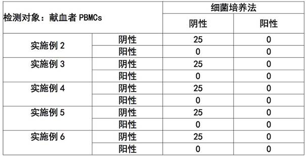
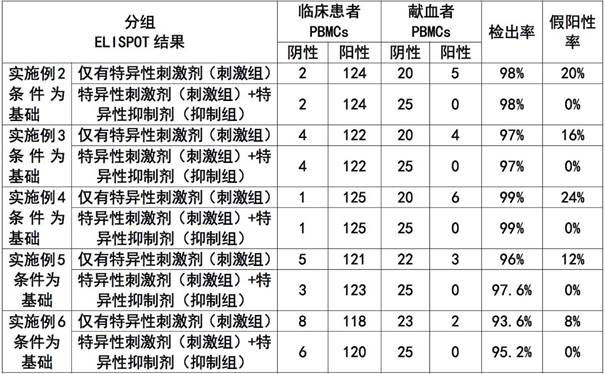
Examples
Embodiment 1
[0046] The preparation of embodiment 1 ELISPOT detection kit
[0047] Specifically, the assembly steps of the ELISPOT detection kit of the present invention are as follows: 96-well filter plate, IFN-γ capture antibody, biotin-labeled IFN-γ detection antibody, specific stimulator, specific inhibitor, negative control (10% 1640 culture medium) and the positive control (phytohemagglutinin PHA) were packaged and assembled into kits respectively.
[0048] Further, the ELISPOT detection kit is assembled as required: enzyme-labeled avidin (avidin-alkaline phosphatase conjugate), NBT / BCIP chromogenic solution, concentrated PBS buffer, blocking solution, cell culture One or more of buffered saline, phosphate buffered saline or phosphate Tween buffered solution.
Embodiment 2
[0049] Example 2 Using the ELISPOT detection kit of Example 1 to detect human peripheral blood blood samples
[0050] The detection method of the present embodiment comprises the following steps:
[0051] 2.1 Dilute the IFN-γ capture antibody to 15 μg / ml with sterile pH 7.4 calcium and magnesium ion-free 1×PBS (ie DPBS) (filtered with a 0.22 μm filter).
[0052] 2.2 Take out the Millipore 96-well PVDF ELISPOT plate (cat: MSIPS4510, Millipore, 96-well filter plate), and add 15 μl of 35% ethanol to each well for 2 minutes.
[0053] 2.3 Wash the filter plate 5 times with sterile water (filtered through a 0.22 μm filter), add 200 μl to each well each time, discard the liquid in the last pass, and gently dry it on sterile paper.
[0054] 2.4 Add 100 μl of the capture antibody solution diluted in step 2.1 to each well, and incubate overnight at 4-8°C.
[0055] 2.5 Remove the antibody coating solution in the filter plate, wash the filter plate 5 times with sterile PBS, 200 μl per w...
Embodiment 3
[0066] Example 3 Using the ELISPOT detection kit of Example 1 to detect human peripheral blood blood samples
[0067] The detection method of the present embodiment comprises the following steps:
[0068] 2.1 Dilute the IFN-γ capture antibody to 15 μg / ml with sterile pH 7.4 calcium and magnesium ion-free 1×PBS (ie DPBS) (filtered with a 0.22 μm filter).
[0069] 2.2 Take out the Millipore 96-well PVDF ELISPOT plate (cat: MSIPS4510, Millipore, 96-well filter plate), and add 15 μl of 35% ethanol to each well for 2 minutes.
[0070] 2.3 Wash the filter plate 5 times with sterile water (filtered through a 0.22 μm filter), add 200 μl to each well each time, discard the liquid in the last pass, and gently dry it on sterile paper.
[0071] 2.4 Add 100 μl of the capture antibody solution diluted in step 2.1 to each well, and incubate overnight at 4-8°C.
[0072] 2.5 Remove the antibody coating solution in the filter plate, wash the filter plate 5 times with sterile PBS, 200 μl per w...
PUM
 Abstract
Description
Claims
Application Information