Method and device for determining arteriovenous diameter ratio in fundus image and electronic equipment

A fundus image, diameter ratio technology, applied in the field of image processing, can solve problems such as difficult to detect changes, complex and laborious

Pending Publication Date: 2020-09-18
北京大恒普信医疗技术有限公司
View PDF2 Cites 1 Cited by
  • Summary
  • Abstract
  • Description
  • Claims
  • Application Information

AI Technical Summary

Problems solved by technology

Arteriolar to venular diameter ratio (AVR) can be used to measure the change in the width of retinal blood vessels, but its relative change is very small, and it is difficult for even experienced ophthalmologists to detect the change in clinical examination, that is, Manual or semi-automatic estimation of AVR values ​​from fundus images is complex and laborious

Method used


Image

  • Method and device for determining arteriovenous diameter ratio in fundus image and electronic equipment
  • Method and device for determining arteriovenous diameter ratio in fundus image and electronic equipment
  • Method and device for determining arteriovenous diameter ratio in fundus image and electronic equipment

Examples


Embodiment approach

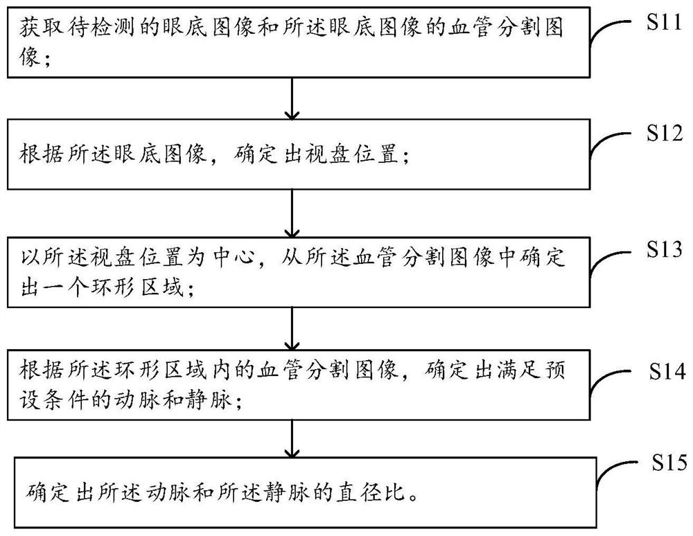
[0052] As an implementation manner, the fundus image to be detected and the blood vessel segmentation image of the fundus image may also be obtained directly from a third party.

[0053] After the fundus image to be detected is acquired, step S12 is executed.

[0054] S12: Determine the position of the optic disc according to the fundus image.

[0055] Since the optic disc is an approximately circular / elliptical highlighted area in the fundus image, generally, there is only one circular / elliptical highlighted area in the fundus image, which is where the optic disc is located. Wherein, the position of the optic disc can be represented by the position of a point at the center of the optic disc, or can be represented by the positions of multiple points representing the contour of the shape and size of the optic disc. According to the pre-established coordinate system in the embodiment of the present application, The blobdetector in opencv is used to determine the position of the...


PUM


Abstract

The invention provides a method and device for determining the arteriovenous diameter ratio in a fundus image and electronic equipment. The method comprises the steps that the fundus image to be detected and a blood vessel segmentation image of the fundus image are acquired; determining the position of an optic disc according to the fundus image; determining an annular region from the blood vesselsegmentation image by taking the optic disc position as a center; determining arteries and veins meeting preset conditions according to the blood vessel segmentation images in the annular area; and determining the diameter ratio of the artery to the vein. An annular region is determined from a blood vessel segmentation image by taking an optic disc position as a center; determining arteries and veins meeting a preset condition by utilizing the blood vessel segmentation image in the annular region; compared with the mode of screening the arteries and veins meeting the preset conditions from the whole blood vessel segmentation image, the calculation complexity of the artery and vein screening process is reduced, manual participation is not needed, and then the artery and vein diameter ratioin the fundus image is automatically determined with low complexity.

Description

technical field [0001] The present application relates to the technical field of image processing, in particular, to a method, device and electronic equipment for determining the ratio of arteriovenous diameters in fundus images. Background technique [0002] Accurately estimating the parameters of retinal vessels has always been a key concern in fundus image analysis. Arteriolar to venular diameter ratio (AVR) can be used to measure the change in the width of retinal blood vessels, but its relative change is very small, and it is difficult for even experienced ophthalmologists to detect the change in clinical examination, that is, Estimating AVR values ​​manually or semi-automatically from fundus images is complex and laborious. Therefore, how to automatically determine the AVR with a relatively low complexity is of great significance. Contents of the invention [0003] In view of this, the purpose of the embodiments of the present application is to provide a method, de...

Claims


Application Information

Patent Timeline
Patent Type & Authority Applications(China)
IPC IPC(8): G06T7/60G06T7/90G06T7/11G06T7/00
CPCG06T7/60G06T7/90G06T7/11G06T7/0012G06T2207/30041G06T2207/30101
Inventor 金蒙唐轶赵雷
Owner 北京大恒普信医疗技术有限公司
Why Eureka
Try Eureka