A method and system for identifying cancer focus regions based on full-slice pathological images
A pathological image and area recognition technology, applied in the field of image processing, can solve complex boundary problems and other problems, achieve good prediction accuracy, simple production method, and improve accuracy
- Summary
- Abstract
- Description
- Claims
- Application Information
AI Technical Summary
Problems solved by technology
Method used
Image
Examples
Embodiment Construction
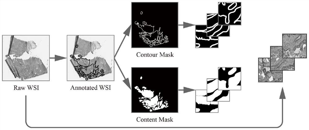
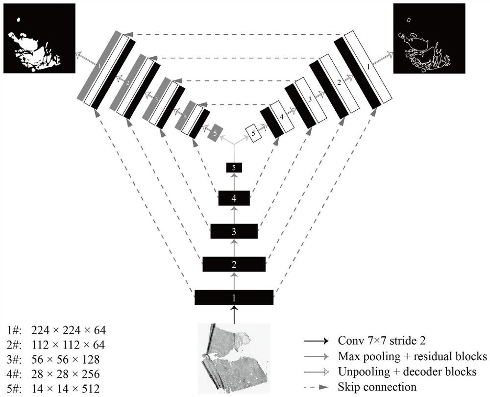
[0045] When we analyzed the characteristics of colorectal cancer pathological images, we found that a very important feature of the cancer focus area is the blurring of the edges. The margins of different subtypes (such as mucinous adenocarcinoma, Injunction cell carcinoma, etc.) have different identification difficulties. Since the cancer focus area is composed of cancer cells, its morphological margins are very complex and have many possibilities, so professional pathologists are often required for identification. Therefore, we propose a new model for this problem, introduce the idea of multi-task learning, and add a parallel contour decoder as a side task on the basis of the improved version of U-Net. In addition to using the mask data of the cancer focus area to supervise the main task, the mask data of the cancer focus outline is also exported to supervise the sub-tasks. In order to strengthen the information fusion of the two tasks, in addition to sharing an encoder, ...
PUM
 Abstract
Description
Claims
Application Information
 - R&D
- Intellectual Property
- Life Sciences
- Materials
- Tech Scout
- Unparalleled Data Quality
- Higher Quality Content
- 60% Fewer Hallucinations
Browse by: Latest US Patents, China's latest patents, Technical Efficacy Thesaurus, Application Domain, Technology Topic, Popular Technical Reports.
© 2025 PatSnap. All rights reserved.Legal|Privacy policy|Modern Slavery Act Transparency Statement|Sitemap|About US| Contact US: help@patsnap.com


