Slice preparation method of non-decalcified osteochondral tissue and osteochondral tissue cell tracing and RNAScope combined test method

A technique for tissue slices and cartilage tissue, which is applied in the joint testing of osteochondral tissue cell tracing and RNAscope, and the preparation of non-decalcified osteochondral tissue slices. Effects of fluorescence interference, avoiding damage and degradation, and saving decalcification time

Pending Publication Date: 2020-12-01
WEST CHINA HOSPITAL SICHUAN UNIV
View PDF6 Cites 1 Cited by
  • Summary
  • Abstract
  • Description
  • Claims
  • Application Information

AI Technical Summary

Benefits of technology

This patented inventions aim to improve the accuracy and speed for studying biomolecules such as calcium (Ca), phosphorus (P), carbohydrates, nucleic acids, enzyme levels, oxidative stress reactions, inflammations caused by external factors like radiation exposure, chemical substances, pathogens, and changes overtime. By embedding these molecular structures into an ice crystal structure instead of freezing them afterwards, this technique allows scientists to examine how cells grow inside their own body while preserving its structural quality. Additionally, there are methods available where certain techniques may use oxygen gas laser irradiating onto the surface layer of calcified areas before making new ones. Overall, these technical improvements make better understanding and exploration of biomedical materials more efficient than current methods.

Problems solved by technology

This patents discusses how fluorochrome-based methods for studying bone growth processes (bone formation) involve combining certain molecules with an agent that specifically targets them during biological activity on the surface of living organisms such as cartilage. These agents may include enzyme catalysts like ribonucleases, lyases, etc., chemical compounds containing amino acids, peptides, proteins, nucleic acid bases, sugar residues, nucleators, antibodies, drugs, small metals, metal chelates, polymers, polysaccharide structures, lipocalites, dyes, cyanines, flavonoid pigments, phthalexine blue, green light absorbing materials, quantum dots, magnetic nanoparticles, iron oxidenanoferritans, calcium carbonate particles, silicon dioxane shell layers, silver nitrate solutions, polyethyleneimine films, and other substances known collectively called stainless steel wires.

Method used


Image

  • Slice preparation method of non-decalcified osteochondral tissue and osteochondral tissue cell tracing and RNAScope combined test method
  • Slice preparation method of non-decalcified osteochondral tissue and osteochondral tissue cell tracing and RNAScope combined test method
  • Slice preparation method of non-decalcified osteochondral tissue and osteochondral tissue cell tracing and RNAScope combined test method

Examples


Embodiment 1

[0055] In this example, 6-week-old model mice were used, which were obtained by crossing Shp2fl / fl mice with Agc1-Cre / ER; Rosa26ZsG mice. The mouse genotype is, Tg(Agc1-CreER; Shp2 fl / + ;Rosa26 ZsG ), the mouse was intraperitoneally injected with tamoxifen within 2 weeks after birth to activate the expression of Cre recombinase, which can only be expressed under the action of Agc1 promoter, and the expression of Cre recombinase can activate the expression of fluorescent protein ZsG, In this way, the cells expressing Agc1 all have the expression of green fluorescent protein ZsG, so that Agc1 positive cells can be traced.

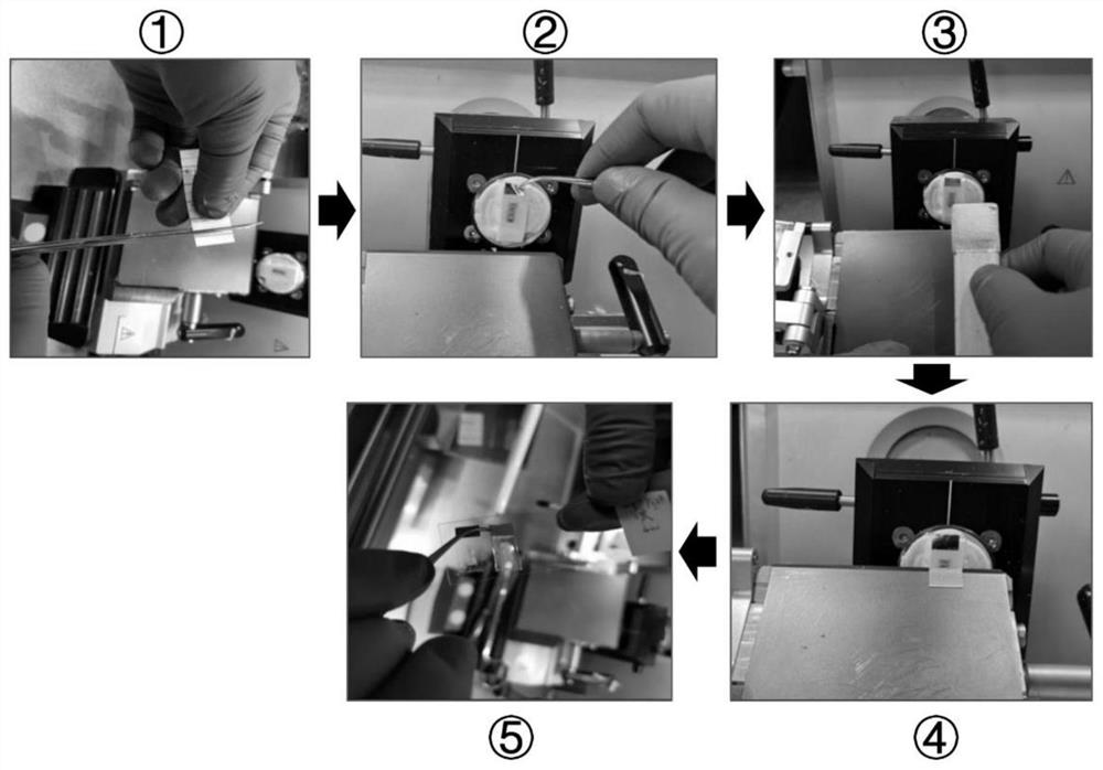
[0056] After preparing the osteochondral tissue slices pasted with Kawamoto membrane in the proximal tibia of the model mouse, RNAscope2.5HD Assay Red Kit produced by Advanced Cell Diagnostics was selected for the in situ hybridization experiment in this example. The process of the in situ hybridization hybridization experiment was performed according to th...

Embodiment 2

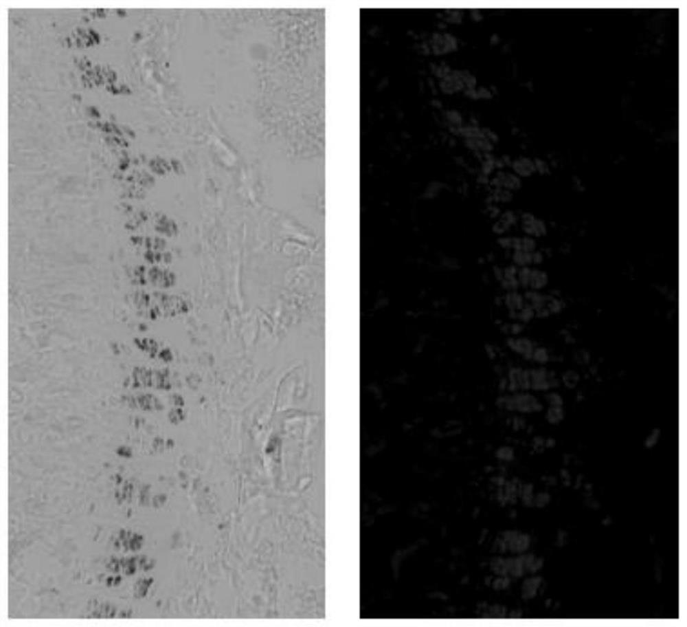
[0058] In this example, the cell tracking method and RNAscope were used to detect the knockout efficiency of the target gene at the mRNA level on the osteochondral tissue section. In this experiment, 10-week-old Tg(Agc1-CreER;Shp2 fl / + ;Rosa26 ZsG ) and Tg(Agc1-CreER; Shp2 fl / fl ;Rosa26 ZsG ) Proximal tibia of model mice. After the osteochondral tissue slices pasted with Kawamoto membrane were prepared, RNAscope 2.5HD Assay RedKit produced by Advanced Cell Diagnostics was used for in situ hybridization experiments. The process of the in situ hybridization hybridization experiment was performed according to the experimental manual of RNAscope 2.5HD Assay RedKit, and each time the osteochondral tissue slice was treated with the reagent, it only needed to cover the osteochondral tissue slice with the reagent. Test results such as Figure 4 shown. Counting the number of red dots among the 100 green cells in the middle region of the growth plate revealed a significant reducti...

Embodiment 3

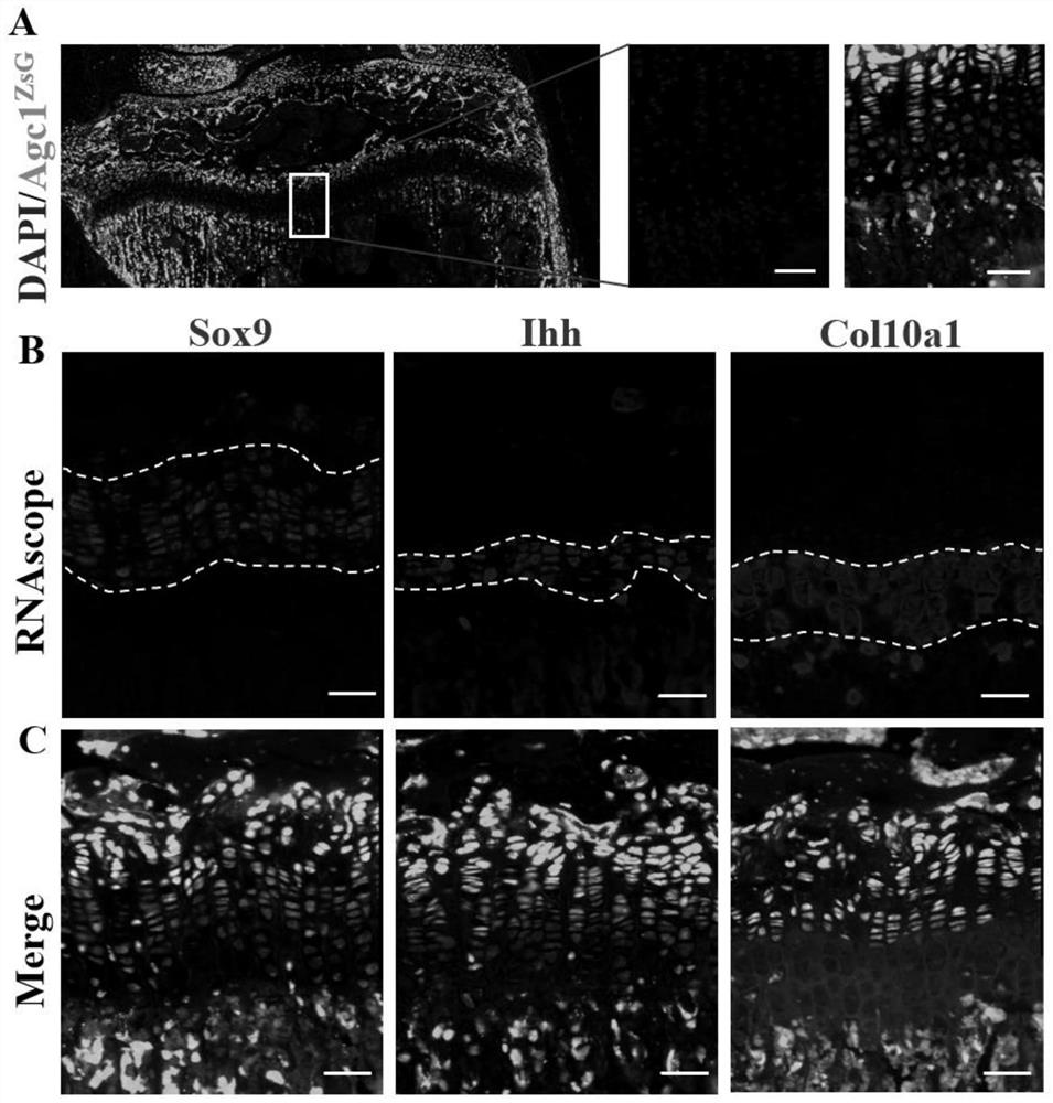
[0060] In this example, the technical solution of the present invention is used to study the pathological changes of osteochondral bone. In this experiment, 10-week-old Tg(Ctsk-Cre;Shp2 fl / fl ;Rosa26 ZsG ) Proximal tibia of model mice. The chondroma on the articular cartilage of the proximal tibia after SHP2 was knocked out in Ctsk-positive cells in mice, the cells of the chondroma originated from Ctsk-positive cells. After the osteochondral tissue slices pasted with Kawamoto membrane were prepared, RNAscope 2.5HD Assay Red Kit produced by Advanced Cell Diagnostics was used for in situ hybridization experiment. The process of the in situ hybridization experiment was performed according to the experimental manual of the RNAscope 2.5HD Assay Red Kit, and each time the osteochondral tissue section was treated with the reagent, it was only necessary to cover the osteochondral tissue section with the reagent. Test results such as Figure 5 shown. In the figure, it can be obser...


PUM


Abstract

The invention belongs to a biological detection technology, and particularly relates to a preparation method of a non-decalcified osteochondral tissue slice and an osteochondral tissue cell tracing and RNAScope combined test method. The problem that in the prior art, due to the fact that a non-decalcified osteochondral tissue slice is difficult to keep complete, the cell tracing and RNAScope combined testing technology cannot be applied to osteochondral tissue slices is solved. The technical scheme of the invention is as follows: the integrity of the osteochondral tissue during frozen sectionis guaranteed through a Kawamoto membrane, and then, a fluorescent cell tracing technology and an in-situ hybridization technology are combined to realize simultaneous observation of a cell tracing fluorescent signal and an RNAsocpe in-situ hybridization specific target RNA signal in the same osteochondral tissue section. The application range of the cell tracing and RNAScope combined test technology is expanded, and the application comprises (1) osteochondral tissue development research, (2) corresponding gene knockout efficiency analysis in osteochondral tissue, (3) research on influences ontranscription levels of other target genes after gene knockout, and (4) osteochondral tissue pathological analysis.

Description


Claims


Application Information

Patent Timeline
Owner WEST CHINA HOSPITAL SICHUAN UNIV
Why Eureka
Try Eureka