A switch-type liposome nano fluorescent probe and its preparation method and application

A nano-fluorescent probe, liposome technology, applied in the field of nanotechnology and fluorescence diagnosis research, can solve the problem of limited switching performance of dye intermolecular distance, etc., and achieve the effects of improving blood stability, improving ACQ efficiency, and reducing interference

Active Publication Date: 2021-06-22
XIDIAN UNIV
View PDF0 Cites 0 Cited by
  • Summary
  • Abstract
  • Description
  • Claims
  • Application Information

AI Technical Summary

Problems solved by technology

However, the existing ACQ-liposome probes are all realized by loading high-concentration hydrophilic dyes in the hydrophilic phase, or encapsulating high-concentration hydrophobic dyes in the hydrophobic phase, and their switching performance is limited by the entrapment. rate and distance between dye molecules

Method used


Image

  • A switch-type liposome nano fluorescent probe and its preparation method and application
  • A switch-type liposome nano fluorescent probe and its preparation method and application
  • A switch-type liposome nano fluorescent probe and its preparation method and application

Examples


Embodiment 1

[0040] The preparation method of DiR-Lipo described in this embodiment is as follows:

[0041] Distearoylphosphatidylcholine (DSPC), cholesterol, distearoylphosphatidylethanolamine-polyethylene glycol (DSPE-PEG2000) in a molar ratio of 55:40:5 (total lipid molar concentration 5mM) Dissolve in chloroform, then remove the chloroform by rotary evaporation to form a lipid film;

[0042] Add 350 mM ammonium sulfate aqueous solution into the lipid film, and rotate in a 60° C. water bath to hydrate the lipid film. After the lipid film to be formed is hydrated to become an emulsion liposome suspension, extrude 10 times on a 100nm polycarbonate film with an extruder;

[0043] The extruded liposome solution was dialyzed 3 times with 100 mM acetate buffer to form a liposome preparation with ammonium sulfate gradient (inside: 350 mM ammonium sulfate; outside: 100 mM acetate buffer)

[0044]Mix the liposome solution obtained in the previous step with the DMSO solution of DiR (DiR account...

Embodiment 2D

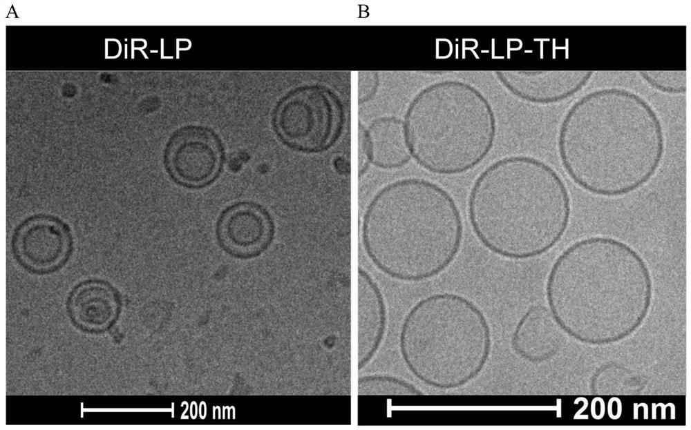
[0049] Morphology comparison of embodiment 2DiR-Lipo and DiR-Lipo-TH

[0050] The DiR-Lipo prepared in Example 1 is indeed encapsulated in the aqueous core of liposomes, and the morphology of DiR-Lipo and DiR-Lipo-TH is characterized by cryo-transmission electron microscopy (Cryo-TEM). The results are shown in figure 2 . Such as figure 2 As shown in A, the DiR-Lipo core exhibits a bilayer conformation with an electron-dense structure, indicating that the ammonium ion forms a complex with DiR and is retained in the aqueous core of the liposome. However, the detection result of DiR-Lipo-TH (2B) did not show a bilayer structure, indicating that DiR entered the hydrophobic phase.

Embodiment 3D

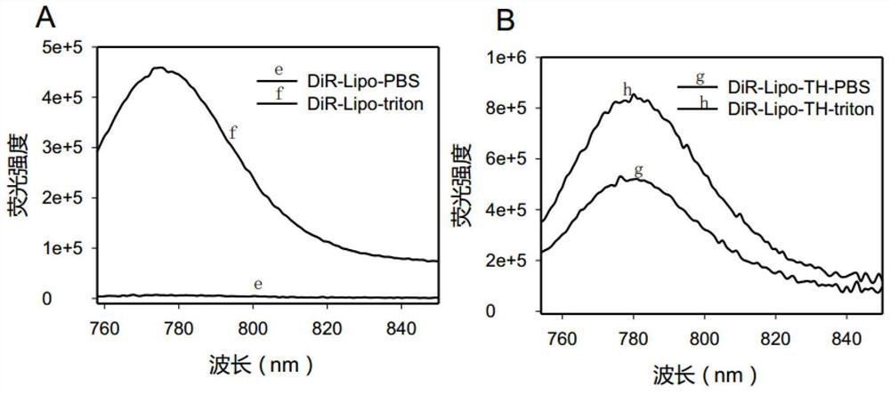
[0051] Example 3 Fluorescence comparison of DiR-Lipo and DiR-Lipo-TH before and after membrane rupture

[0052] The DiR-Lipo prepared in Example 1 is indeed encapsulated in the aqueous core of liposomes, and has in vitro fluorescence activatable properties. This example will use 0.05% Triton X-100 to treat DiR-Lipo and DiR-Lipo -TH perform membrane rupture respectively. First dilute DiR-Lipo and DiR-Lipo-TH with PBS to measure the fluorescence intensity of liposomes in the intact state, then add 0.05% Triton X-100 to disrupt the membrane of liposomes, and measure the dye DiR in monomeric state Fluorescence intensity, compare the fluorescence intensity before and after membrane rupture, the results are shown in image 3 . Depend on image 3 A shows that the fluorescence intensity of DiR-Lipo after membrane rupture (f) is much greater than the fluorescence intensity before membrane rupture (e), and the fluorescence enhancement factor is 70 times, indicating that the fluoresce...


PUM


Abstract

A switch-type liposome nanometer fluorescent probe comprises a near-infrared fluorescent dye and a liposome phospholipid membrane wrapped outside it; the liposome phospholipid membrane is composed of choline derivatives, cholesterol and distearoylphosphatidylethanolamine- Composed of polyethylene glycol 2000; the switch-type liposome nano-fluorescent probe encapsulates the hydrophobic dye in the liposome inner core, and compared with the traditional method of loading the hydrophobic dye on the lipid bilayer, the loading rate of the dye is higher High, better stability, and combined with the advantages of fluorescence can be activated. The preparation process of the probe is simple, the cost is low, the use is convenient, and the imaging effect in the body is good.

Description

technical field [0001] The invention belongs to the field of nanotechnology and fluorescent diagnosis research, and in particular relates to a switch-type liposome nanometer fluorescent probe and its preparation method and application. Background technique [0002] The near-infrared fluorescence imaging technology for tumors has excellent resolution and sensitivity, and the penetration depth of near-infrared fluorescence imaging in biological tissues is large, and the imaging signal is less affected by the tissue. It is a kind developed in recent years. new imaging methods. Fluorescent contrast agents (such as indocyanine green (ICG), methylene blue (MB), etc.) themselves have poor tumor selectivity, resulting in a very low tumor signal-to-background ratio (TBR). TBR is considered to be an important criterion for evaluating and selecting fluorescent contrast agents, so improving the TBR of fluorescent probes is very important for tumor fluorescence imaging. Encapsulating t...

Claims


Application Information

Patent Timeline
Patent Type & Authority Patents(China)
IPC IPC(8): A61K49/00
CPCA61K49/0032A61K49/0084
Inventor 夏玉琼许春仲张象涵王忠良张瑞丽吴延坤李采儒
Owner XIDIAN UNIV
Why Eureka
Try Eureka