Retinal vessel segmentation method in fundus image and computer readable storage medium

A technology of retinal blood vessels and fundus images, which is applied in the field of medical image processing to achieve good segmentation performance

Inactive Publication Date: 2021-01-15
SHENZHEN GRADUATE SCHOOL TSINGHUA UNIV
View PDF2 Cites 30 Cited by
  • Summary
  • Abstract
  • Description
  • Claims
  • Application Information

AI Technical Summary

Problems solved by technology

[0006] In order to solve the existing problems, the present invention provides a method for segmenting retinal blood vessels in fundus images and a computer-readable storage medium

Method used


Image

  • Retinal vessel segmentation method in fundus image and computer readable storage medium
  • Retinal vessel segmentation method in fundus image and computer readable storage medium
  • Retinal vessel segmentation method in fundus image and computer readable storage medium

Examples


Embodiment Construction

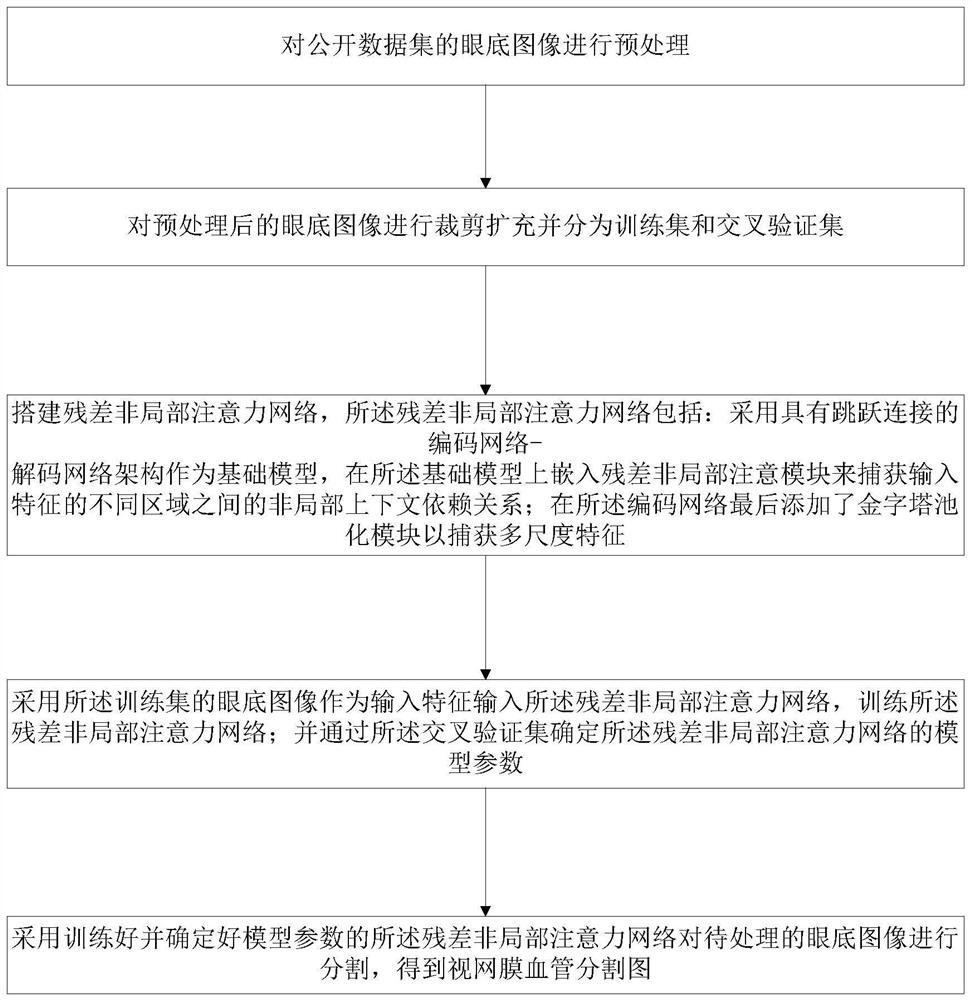
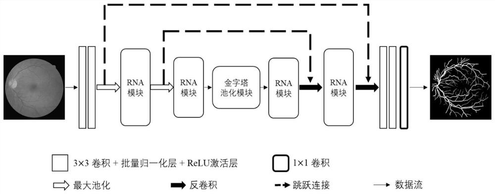
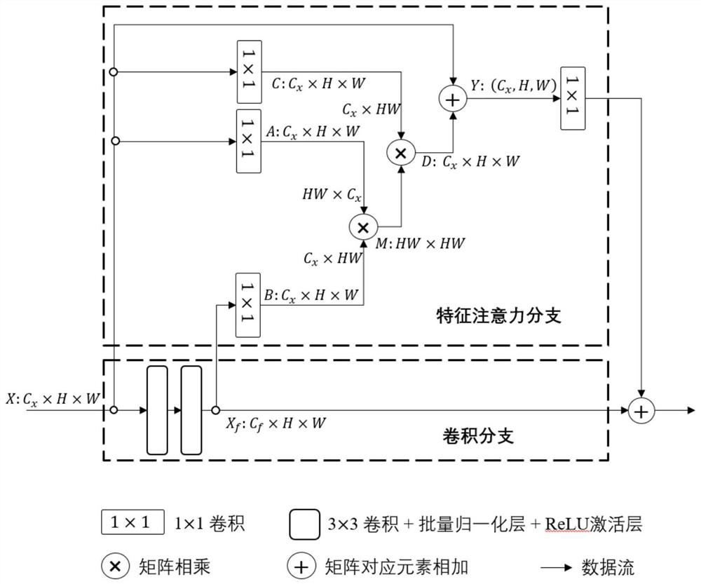
[0030]In order to make the technical problems, technical solutions, and beneficial effects to be solved by the embodiments of the present invention clearer, the following further describes the present invention in detail with reference to the accompanying drawings and embodiments. It should be understood that the specific embodiments described herein are only used to explain the present invention, but not to limit the present invention.

[0031]It should be noted that when an element is referred to as being "fixed to" or "disposed on" another element, it can be directly on the other element or indirectly on the other element. When an element is said to be "connected to" another element, it can be directly connected to the other element or indirectly connected to the other element. In addition, the connection can be used for fixing or circuit connection.

[0032]It should be understood that the terms "length", "width", "upper", "lower", "front", "rear", "left", "right", "vertical", "horizo...


PUM


Abstract

The invention provides a retinal vessel segmentation method in a fundus image and a computer readable storage medium. The method comprises the steps of preprocessing the fundus image of a public dataset; cutting and expanding the preprocessed fundus image and dividing the fundus image into a training set and a cross validation set; building a residual non-local attention network, wherein the residual non-local attention network comprises the steps of adopting a coding network decoding network architecture with jump connection as a basic model, and embedding a residual non-local attention module to capture a non-local context dependency relationship between different areas of input features; adding a pyramid pooling module to the encoding network to capture multi-scale features; training aresidual non-local attention network by adopting the training set; determining model parameters of the residual non-local attention network through the cross validation set; and adopting the trainedresidual non-local attention network to segment the fundus image to be processed, so that a retinal vessel segmentation image can be obtained. Segmentation performance is better.

Description

Technical field[0001]The present invention relates to the technical field of medical image processing, in particular to a method for segmenting retinal blood vessels in fundus images and a computer-readable storage medium.Background technique[0002]The analysis of retinal blood vessels can provide rich information about eye conditions and human health. Among them, obtaining a clear blood vessel distribution map is a crucial step.[0003]Recently, retinal blood vessel segmentation methods often use Convolutional Neural Network (CNN) as the basic model algorithm, and this model algorithm has shown excellent feature extraction capabilities in segmentation tasks. However, the fixed receptive field prevents the network from adapting to changes in the scale of retinal blood vessels. Because the retinal vessel structure is complex, entangled and multi-scale. In addition, the local receptive field cannot capture the global information and context dependence between different regions of the ent...

Claims


Application Information

Patent Timeline
Patent Type & Authority Applications(China)
IPC IPC(8): G06T7/12G06K9/62G06T3/40G06N3/04
CPCG06T7/12G06T3/4007G06T2207/20081G06T2207/30041G06N3/045G06F18/2415G06F18/214
Inventor 张凯陈艺璇董宇涵
Owner SHENZHEN GRADUATE SCHOOL TSINGHUA UNIV
Why Eureka
Try Eureka