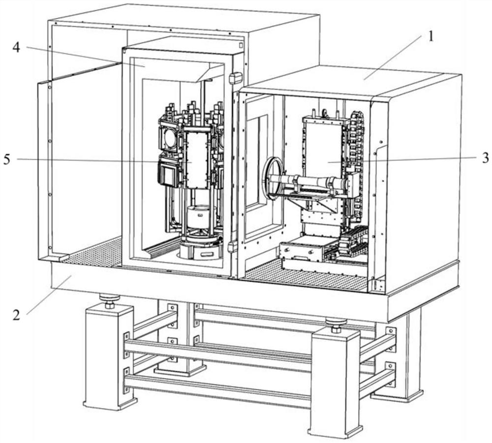
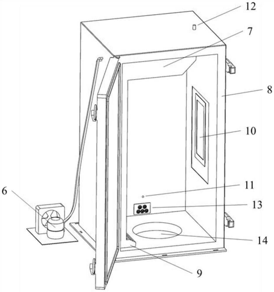
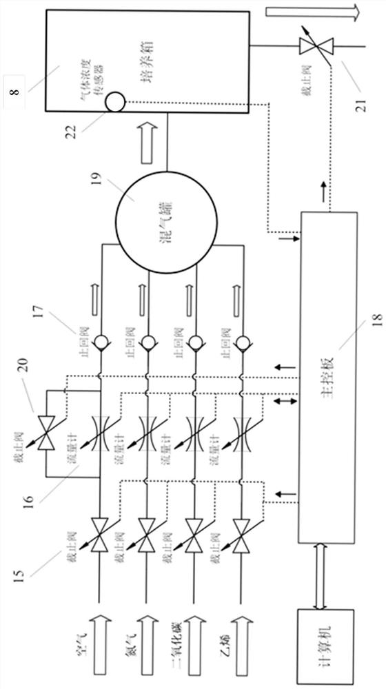
Biological sample imaging equipment
A technology of biological samples and imaging equipment, which is applied in the field of biological instruments, can solve the problems of inability to conveniently control plant growth conditions and meet the needs of scientific research scenarios, and achieve the effect of reducing interference
- Summary
- Abstract
- Description
- Claims
- Application Information
AI Technical Summary
Problems solved by technology
Method used
Image
Examples
example 1
[0340] Example 1: Studying cabbage seed germination and seedling growth using a large horizontal petri dish stand 1. Sterilization of the petri dish: disassemble the parts of the petri dish, wash with detergent and water, rinse three times with distilled water, and dry at room temperature; Install the sealing strip in the groove of the tank and fix the front cover with screws, and seal it with the top cover into a paper sealing bag. After sterilized by ethylene oxide gas, disassemble the paper bag in the ultra-clean workbench and take it out for use;
[0341] 2. Sterilize the surface of cabbage seeds: put an appropriate amount of seeds in a 1.5mL centrifuge tube, soak in 75% alcohol + 0.01% Triton X-100 and shake fully for 15 minutes, pour off the liquid; add 95% alcohol to rinse once, pour off the liquid ;Tighten the tube cap after air-drying thoroughly in the ultra-clean workbench;
[0342]3. Prepare medium: 4.33g / L Murashige-Skoog salt, 10g / L sucrose, 3g / L Phytagel plant ge...
example 2
[0348] Example 2: An example of microscopic phenotyping to study the dynamic details and mechanism of shade avoidance responses of Arabidopsis seedlings
[0349] 1. Imaging module installation: install the microscopic phenotype detection module on the dovetail guide rail 76 of the three-dimensional displacement control module, and lock it through the positioning hole 77; the lens of the microscopic phenotype detection module uses a 4-fold magnified bi-telecentric lens;
[0350] 2. Installation of the plant cultivation module: install 16 standard small vertical culture dish racks 36 on the frame fixing parts 35 of the eight-way main bracket; insert the wiring of the top cultivation light source 44 into the electrical socket interface 34, and the top cultivation light source Use a three-in-one LED lamp bead with three monochromatic lights: 660nm red light, 735nm far-red light, and 450nm blue light;
[0351] 3. Seed sterilization: the same as the seed sterilization method introdu...
example 3
[0362] Example 3: An example of microscopic phenotyping to study the rapid response of Arabidopsis seedlings to ethylene growth inhibition
[0363] 1. Imaging module installation, seed sterilization and medium configuration are the same as Example 2;
[0364] 2. Plant sample preparation: Use tweezers to sow sterilized seeds on the surface of the culture medium in an ultra-clean workbench, and arrange them in a row with an interval of about 5mm; multiple rows can be sown in each petri dish, with an interval of 20mm between each row; cover the petri dish , use a parafilm to seal; place it in the dark at 4°C after imbibition for 4 days, take it out and wrap it in aluminum foil, place it vertically at 22°C in the dark and culture it for 2 days, the Arabidopsis seedlings should be close to the surface of the medium and vertical Growth; keep only dim green LED bulbs for illumination in a completely dark room, open the petri dish in the ultra-clean workbench, select plants in good gr...
PUM
| Property | Measurement | Unit |
|---|---|---|
| thickness | aaaaa | aaaaa |
Abstract
Description
Claims
Application Information
 - R&D
- Intellectual Property
- Life Sciences
- Materials
- Tech Scout
- Unparalleled Data Quality
- Higher Quality Content
- 60% Fewer Hallucinations
Browse by: Latest US Patents, China's latest patents, Technical Efficacy Thesaurus, Application Domain, Technology Topic, Popular Technical Reports.
© 2025 PatSnap. All rights reserved.Legal|Privacy policy|Modern Slavery Act Transparency Statement|Sitemap|About US| Contact US: help@patsnap.com


