Method for constructing lung adenocarcinoma infiltration imaging omics classification model
A radiomics and classification model technology, applied in image analysis, image enhancement, image data processing, etc., can solve the problems of poor generalization performance of radiomics models, dependence on invasive operations, lack of accuracy and stability, etc. , to achieve the effect of solving the problem of low generalization or robustness, saving labor costs, and ensuring accuracy and robustness
Pending Publication Date: 2021-03-26
SHANGHAI CHEST HOSPITAL
View PDF0 Cites 0 Cited by
- Summary
- Abstract
- Description
- Claims
- Application Information
AI Technical Summary
Problems solved by technology
[0006] The purpose of the present invention is to use multi-resolution CT images of the same patient to construct a radiomics classification model of lung adenocarcinoma invasion with good robustness, high prediction accuracy and non-invasiveness, which is suitable for
Method used
View moreImage
Smart ImageViewing Examples

Examples
 PUM
 Abstract
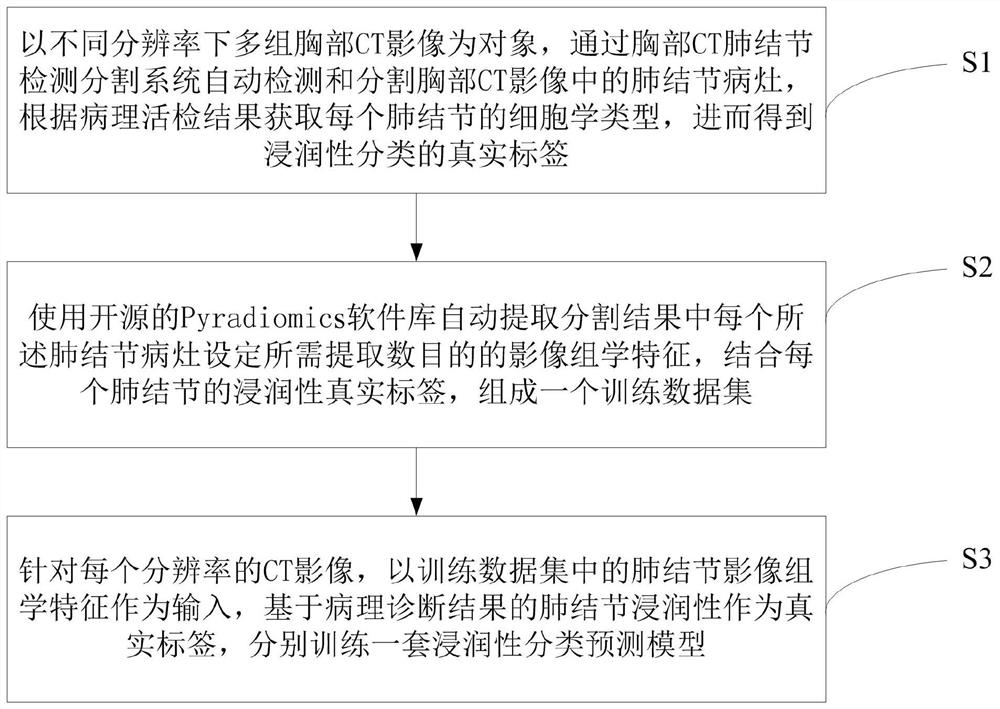
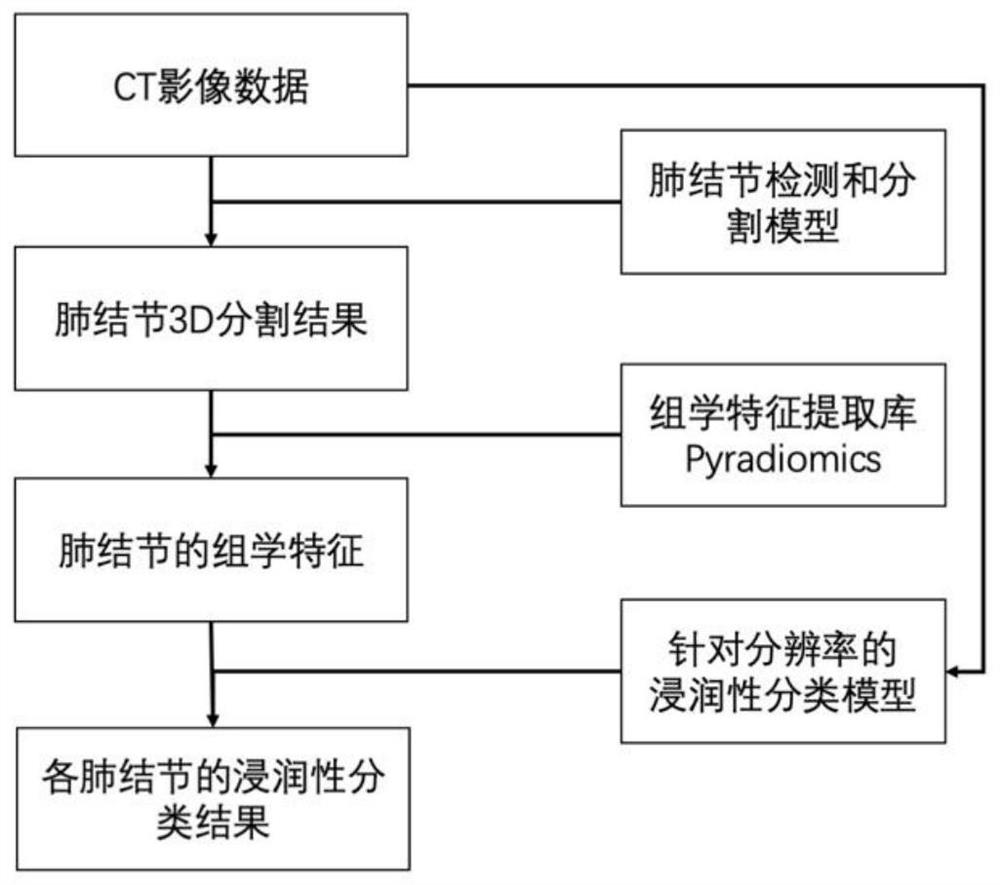
The invention discloses a method for constructing lung adenocarcinoma infiltration imaging omics classification model, which comprises the following steps: by taking multiple groups of chest CT imagesunder different resolutions as objects, automatically detecting and segmenting pulmonary nodule lesions in the chest CT images through a chest CT pulmonary nodule detection and segmentation system, and obtaining a cytology type of each pulmonary nodule according to a pathological biopsy result, and obtaining a real label of wettability classification; using an open-source Pyraliomics software library to automatically extract a required extraction number of image omics features set for each pulmonary nodule lesion in the segmentation result, and forming a training data set in combination withthe wettability real label of each pulmonary nodule; aiming at the CT images with each resolution, respectively training a set of wettability classification prediction model by taking pulmonary noduleimage omics characteristics in the training data set as input and pulmonary nodule wettability based on a pathological diagnosis result as a real label. According to the construction method, a seriesof optimal models are obtained, and the generalization performance of the models on CT with different resolutions is guaranteed.
Description
technical field [0001] The invention belongs to the technical field of medical data processing, and in particular relates to a method for constructing a radiomics classification model of invasive lung adenocarcinoma. Background technique [0002] The differential diagnosis of invasiveness of early tumors is the key to preoperative evaluation and determines the follow-up clinical treatment. For example, pathologically confirmed invasive early lung adenocarcinoma has a poor prognosis and requires total lobectomy; non-invasive early lung adenocarcinoma (including atypical adenomatous hyperplasia, carcinoma in situ, and minimally invasive carcinoma) has a better prognosis. Observation or partial lobectomy may be performed. However, the current pathological diagnosis requires the use of invasive operations to obtain lesion tissue, including puncture, thoracoscopic surgery, and thoracotomy, and the final pathological results may prove that these invasive operations are unnecessar...
Claims
Login to View More Application Information
Patent Timeline
 IPC IPC(8): G06K9/62G06T7/00G06T7/11
CPCG06T7/0012G06T7/11G06T2207/30064G06T2207/10081G06T2207/20064G06T2207/30096G06F18/241G06F18/214
Inventor 陶广昱于红张晓军尹乐康陈怡楠叶晓丹虞凌明郑志春施珏倩石德军周振
Owner SHANGHAI CHEST HOSPITAL