
Deep brain tumor and white matter fiber bundle relation model and preparation method thereof

A white matter fiber and relational model technology, applied in the field of medical models, can solve problems such as spatial imagination, three-dimensional space understanding difficulties, and limited visualization, so as to reduce neurological disability, increase effectiveness, and reduce surgical complications. Effect

Active Publication Date: 2021-03-26
天津市第一中心医院
View PDF3 Cites 1 Cited by
  • Summary
  • Abstract
  • Description
  • Claims
  • Application Information

AI Technical Summary

Problems solved by technology

[0003] In clinical practice, there are individual differences in the course of the subcortical conduction pathway-white matter fiber bundles. The white matter fibers around the tumor are pushed, squeezed, interrupted and destroyed, and the three-dimensional relationship between the tumor and nerve fibers is limited in visualization. , Clinically, different neurosurgeons and medical students have different spatial imaginations, and the existence of influencing factors such as difficulty in understanding the three-dimensional space of the structure of the surgical area only on the two-dimensional level has led to accurate preoperative evaluation of deep brain tumors, Accurate implementation of intraoperative tumor resection and effective medical surgery training and medical teaching practice face certain difficulties and challenges

Method used


Image

  • Deep brain tumor and white matter fiber bundle relation model and preparation method thereof
  • Deep brain tumor and white matter fiber bundle relation model and preparation method thereof
  • Deep brain tumor and white matter fiber bundle relation model and preparation method thereof

Examples


Embodiment Construction

[0040] The technical solutions of the present invention will be clearly and completely described below in conjunction with the accompanying drawings. Apparently, the described embodiments are some of the embodiments of the present invention, but not all of them. Based on the embodiments of the present invention, all other embodiments obtained by persons of ordinary skill in the art without making creative efforts belong to the protection scope of the present invention.

[0041] In the description of the present invention, it should be noted that the terms "center", "upper", "lower", "left", "right", "vertical", "horizontal", "inner", "outer" etc. The indicated orientation or positional relationship is based on the orientation or positional relationship shown in the drawings, and is only for the convenience of describing the present invention and simplifying the description, rather than indicating or implying that the referred device or element must have a specific orientation, ...


PUM


Abstract

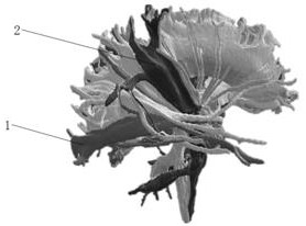
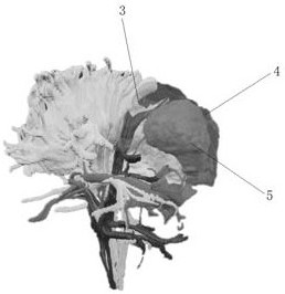
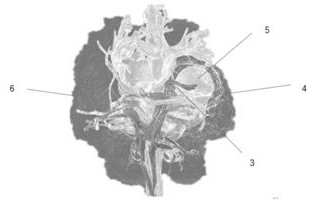
The invention provides a deep brain tumor and white matter fiber bundle relation model and a preparation method thereof, and aims to obtain 3D high-resolution scanning images of deep brain tumors, adjacent white matter fiber bundles and brain parenchyma through an MR multi-modal imaging sequence. The obtained original image data are input into modeling post-processing software, and image fusion, segmentation and reconstruction are carried out on deep brain tumors, internal hemorrhage, peripheral edema, adjacent functional fiber bundles and brain parenchyma structures to obtain a deep brain tumor three-dimensional anatomical model; and the model can be imported into a color 3D printer to print an individualized precise medical 3D printing solid model for the brain tumor. The front-and-backand up-and-down spatial relationship between white matter fiber bundles such as brain tumor and adjacent key visual bundles, cortical spinal cord bundles and corpus callosum and brain parenchyma is accurately positioned. The occurrence probability of postoperative visual and dyskinesia is reduced, and the requirements of precise medical evaluation and operation are met. The model is used for preoperative planning and operation simulation, serves as a precise medical teaching training model, and improves the medical training teaching effect.

Description

technical field [0001] The invention belongs to the technical field of medical models, in particular to a model of the relationship between deep brain tumors and white matter fiber bundles and a preparation method thereof. Background technique [0002] Traditional microsurgery for deep brain tumors has a low total resection rate and a high morbidity rate, and postoperative permanent neurological dysfunction is likely to occur. How to remove the tumor safely and accurately to the greatest extent is particularly important. Deep brain tumors are more common in glioma, metastatic tumor, lymphoma, etc. Since the tumor is located in the deep brain parenchyma and deep in the cortex of the functional area, the risk of surgery is high; how to remove the tumor accurately, safely, quickly and completely has always troubled the nerves. surgeon. [0003] In clinical practice, there are individual differences in the course of the subcortical conduction pathway-white matter fiber bundles....

Claims


Application Information

Patent Timeline
IPC IPC(8): G06T17/00G06T5/50G06T7/11G06T7/136B29C64/386B33Y50/00
CPCG06T17/00G06T5/50G06T7/11G06T7/136B29C64/386B33Y50/00G06T2207/10016G06T2207/20221G06T2207/30016G06T2207/30096
Inventor 刘丽华秦文丁浩王国鹤于春水
Owner 天津市第一中心医院
Learn More