Tissue elasticity detection method, device and system

An elasticity detection and organization technology, applied in the field of measurement, can solve problems such as inaccuracy, unstable imaging results or measurement results, and achieve the effect of improving stability and accuracy and improving operation efficiency.

Pending Publication Date: 2021-06-22
WUXI HISKY MEDICAL TECH +1
View PDF9 Cites 1 Cited by
  • Summary
  • Abstract
  • Description
  • Claims
  • Application Information

AI Technical Summary

Problems solved by technology

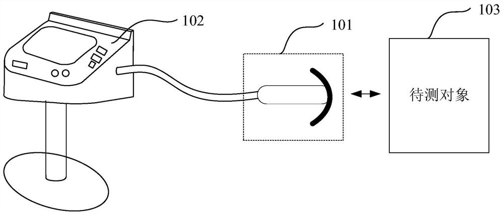
[0005] Embodiments of the present invention provide a tissue elasticity detection method, device, and system to solve the problem caused by ultrasonic imaging and elastic measurement in the

Method used


Image

  • Tissue elasticity detection method, device and system
  • Tissue elasticity detection method, device and system
  • Tissue elasticity detection method, device and system

Examples


Embodiment Construction

[0041] In order to make the purpose, technical solutions and advantages of the embodiments of the present invention clearer, the technical solutions in the embodiments of the present invention will be clearly and completely described below in conjunction with the drawings in the embodiments of the present invention. Obviously, the described embodiments It is only some embodiments of the present invention, but not all embodiments. Based on the embodiments of the present invention, all other embodiments obtained by persons of ordinary skill in the art without making creative efforts belong to the protection scope of the present invention.

[0042] The terms "first", "second", "third", "fourth", etc. (if any) in the description and claims of the present invention and the above drawings are used to distinguish similar objects and not necessarily Describe a specific order or sequence. It is to be understood that the data so used are interchangeable under appropriate circumstances ...


PUM


Abstract

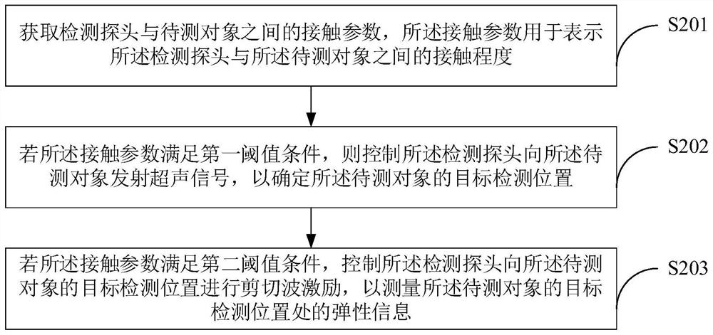
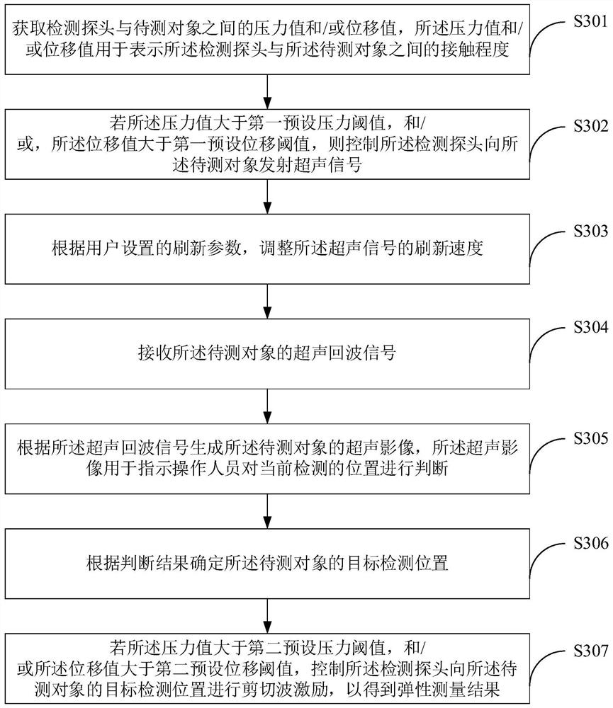
The embodiment of the invention provides a tissue elasticity detection method, device and system. The method comprises the steps: obtaining a contact parameter between a detection probe and a to-be-detected object, the contact parameter being used for representing the contact degree between the detection probe and the to-be-detected object; if the contact parameter meets a first threshold condition, controlling the detection probe to transmit and receive an ultrasonic signal to the to-be-detected object so as to determine a target detection position of the to-be-detected object; and if the contact parameter meets a second threshold condition, controlling the detection probe to perform shear wave excitation on the target detection position of the to-be-detected object so as to measure the elasticity of the target detection position of the to-be-detected object. According to the embodiment of the invention, an operator can accurately judge the opportunity of ultrasonic signal emission and shear wave excitation, so that the stability and accuracy of a measurement result are improved.

Description

technical field [0001] Embodiments of the present invention relate to the field of measurement technology, and in particular to a tissue elasticity detection method, device and system. Background technique [0002] Ultrasound imaging and elastography are widely used in medical care and other fields. Usually, the detection probe is used to emit ultrasonic signals (such as: A-ultrasound, M-ultrasound, or B-ultrasound signals) for ultrasonic image guidance to initially determine whether the current detection position is the detection object The position and whether there is interference, after obtaining the position of the detection object, perform shear wave excitation (such as: mechanical wave or shear wave signal), and measure the elasticity of the detection object. Combining the above two detection methods can realize real-time Quickly obtain the position of the detection object, and obtain the elasticity of the detection object through elasticity measurement. [0003] In ...

Claims


Application Information

Patent Timeline
IPC IPC(8): A61B8/08A61B8/00
CPCA61B8/085A61B8/485A61B8/4444A61B8/54
Inventor 何琼邵金华孙锦
Owner WUXI HISKY MEDICAL TECH
Why Eureka
Try Eureka