Auxiliary film for vascular catheterization operation

A film and surgery technology, applied in the medical field, can solve the problems of increasing the preparation time of ultrasound probes and increasing the fear of patients

Pending Publication Date: 2021-08-20
CHONGQING UNIV CANCER HOSPITAL
View PDF0 Cites 0 Cited by
  • Summary
  • Abstract
  • Description
  • Claims
  • Application Information

AI Technical Summary

Problems solved by technology

[0005] However, before each operation, two surgical sutures must be tied to the ultrasound probe, and the sterile film is used to fix the double sutures, and then the catheterization operation is performed, which increases the preparation time of the ultrasound probe and increases the patient's time during the operation. previous fear

Method used


Image

  • Auxiliary film for vascular catheterization operation
  • Auxiliary film for vascular catheterization operation

Examples


Embodiment 1

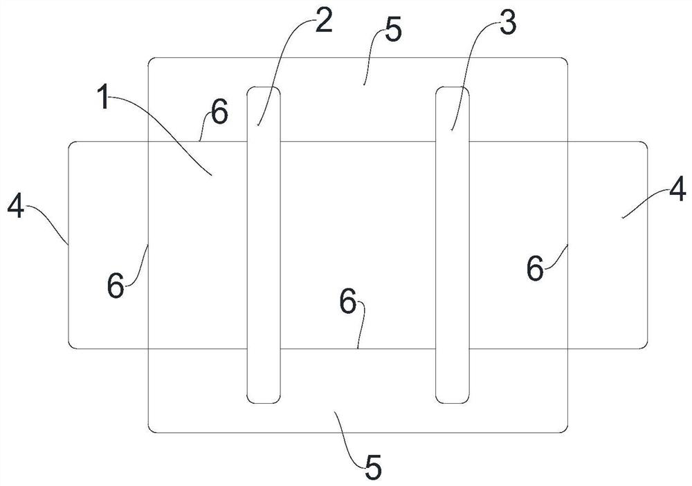
[0030] Such as figure 1 As shown, this embodiment provides an auxiliary patch for vascular catheterization surgery. The auxiliary patch includes a sterile sticking part 1 that can penetrate ultrasonic waves and a first barrier 2 and a second barrier that prevent ultrasound from penetrating. The above-mentioned first blocking piece 2 and the above-mentioned second blocking piece 3 are arranged on the above-mentioned adhesive piece 1 , and there is a gap between the first blocking piece 2 and the second blocking piece 3 .

[0031] In this embodiment, the auxiliary film is a whole, and the first blocking piece 2 and the second blocking piece 3 are arranged on the sticking piece 1, and there is a gap between the first blocking piece 2 and the second blocking piece 3, and the tube insertion operation is performed. It is simple and convenient, and it greatly reduces the preparation time before the catheterization operation, reduces the waiting time of the patient, and correspondingl...

Embodiment 2

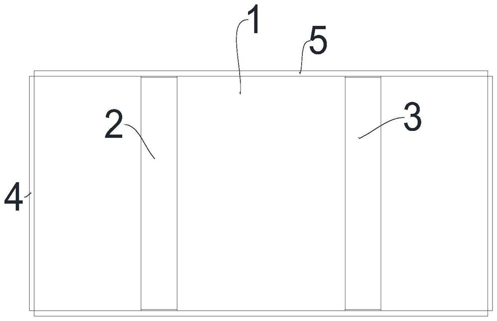
[0034] Such as figure 1 , 2 As shown, this embodiment is based on the above-mentioned embodiment 1, and the above-mentioned first blocking member 2 and the above-mentioned second blocking member 3 are parallel.

[0035] In this embodiment, the above-mentioned first stopper 2 and the above-mentioned second stopper 3 are parallel, and when the adhesive part 1 is bonded on the ultrasonic probe, the first stopper 2 and the second stopper 3 should be aligned with the long axis of the ultrasonic probe. It is vertical to ensure that after the puncture needle penetrates into the skin, the observation area of ​​the operator is larger and more intuitive, and the active area of ​​the puncture needle is larger.

[0036] The distance between the first stopper 2 and the second stopper 3 is a certain distance. The transverse diameter of the internal jugular vein in adult internal jugular vein catheterization surgery is about 12mm-15mm. The distance between the first stopper 2 and the second...

Embodiment 3

[0038] Such as figure 1 , 2 As shown, this embodiment is based on some of the above-mentioned embodiments, and the area of ​​the above-mentioned sticker 1 is larger than the area of ​​the ultrasonic probe.

[0039] In this embodiment, the area of ​​the sticking part 1 is larger than the area of ​​the ultrasonic probe to ensure that the sticking part 1 can completely cover the ultrasonic probe. Contaminants coming into contact with the skin of the opening can cause the opening to become infected, leading to other medical conditions in the patient.

[0040] Such as figure 1 , 2 As shown, in some implementations of this embodiment, the above-mentioned sticker 1 is provided with creases 6 .

[0041] In the above-mentioned embodiment, the adhesive member 1 is provided with creases 6, that is, there are many creases 6, and the area surrounded by the multiple creases 6 is the same as the area of ​​the front end of the ultrasonic probe. When the adhesive member 1 is installed , you...


PUM


Abstract

The invention provides an auxiliary film for a vascular catheterization operation, and relates to the technical field of medical treatment. The auxiliary film for the vascular catheterization operation comprises a sterile sticking piece capable of being penetrated by ultrasonic waves, a first blocking piece and a second blocking piece, wherein the first blocking piece and the second blocking piece are used for preventing the ultrasonic waves from penetrating through the sticking piece, are arranged on the sticking piece and can be directly stuck on an ultrasonic probe, so that the preoperative preparation time is short; and the first blocking piece and the second blocking piece are convenient for guiding a puncture needle to select a puncture position and helping the puncture needle to be stably and safely inserted into a blood vessel, so that accurate and safe intubation is realized, and the generation of mechanical complications is reduced.

Description

technical field [0001] The invention relates to the field of medical technology, in particular to an auxiliary patch used in vascular catheterization operations. Background technique [0002] Real-time ultrasound-guided vascular (arterial or venous) cannulation has become a standard surgical approach with higher success rates and fewer mechanical complications than anatomically landmarked techniques. At present, the commonly used methods for ultrasound-guided vascular cannulation include short-axis out-of-plane and long-axis in-plane. [0003] However, both the short-axis out-of-plane technique and the long-axis in-plane technique have major disadvantages. The short-axis out-of-plane technique and the long-axis in-plane technique are used independently, and the observation range is narrow, and the puncture needle is easy to puncture other tissues by mistake, resulting in Numerous mechanical complications occurred. [0004] We propose an improved combination of short and lo...

Claims


Application Information

Patent Timeline
Patent Type & Authority Applications(China)
IPC IPC(8): A61B8/00A61B17/34
CPCA61B8/4422A61B8/4444A61B17/3403A61B2017/3413
Inventor 唐家喜王玲赵娟刘红亮辇伟奇唐万燕
Owner CHONGQING UNIV CANCER HOSPITAL
Why Eureka
Try Eureka