CTA three-dimensional reconstruction mirror image data image projection method, image processing method and device
A technology of mirror data and three-dimensional reconstruction, applied in the field of medical image processing, can solve the problems of discussing the essential difference between CAG and CTA, difficult for PCI operators to understand and accept, complicated operation process, etc., to achieve convenient treatment plan, reduce the difficulty of interventional surgery, The effect of correct spatial relationship
- Summary
- Abstract
- Description
- Claims
- Application Information
AI Technical Summary
Problems solved by technology
Method used
Image

Examples
no. 1 example
[0057] The CTA three-dimensional fusion method capable of precise fusion with angiography according to the first embodiment of the present invention includes the following steps.
[0058] S1: Obtain 3D reconstruction image data
[0059] Import the 3D reconstruction image data obtained from CT scanning, such as CTA 3D data, into UG NX11.0 or other 3D software. Thus, the CTA three-dimensional data of the spine and the CTA three-dimensional data of the location of the lesion can be obtained. The CTA three-dimensional data of the spine, thoracic spine, ribs, etc. are used as reference positions for the position of the CTA three-dimensional data of the lesion, but the spine is given priority. In other words, the relative position of the lesion (relative to the position of the spine) is the key position information for reconstruction. Here, the lesion may be the heart, or the kidney, or the like.
[0060] S2: Set the image projection mode of CTA 3D reconstruction mirror data to p...
no. 2 example
[0090] The CTA three-dimensional fusion method for precise fusion with angiography in the second embodiment of the present invention includes the following steps.
[0091] S1: Obtain 3D reconstruction image data
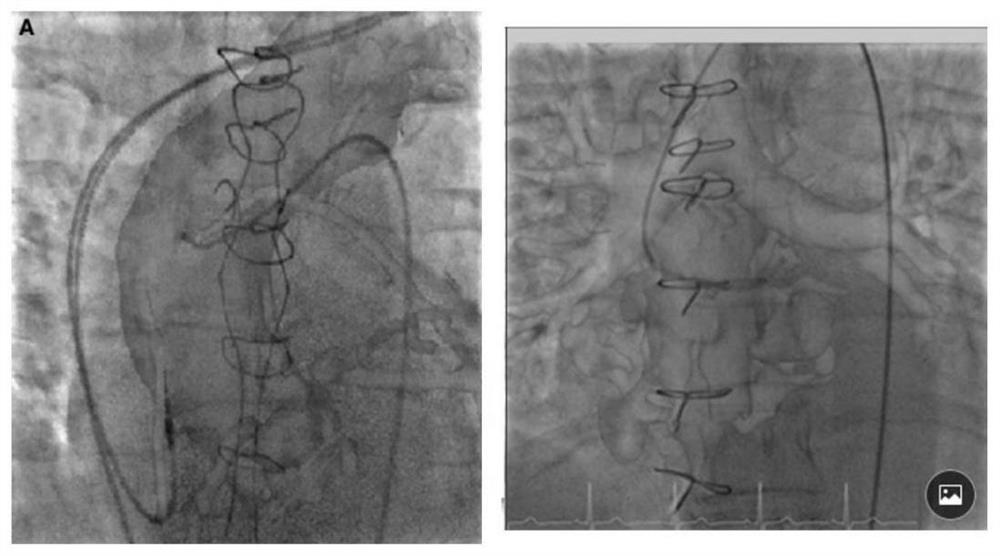
[0092] Similar to the first embodiment, the three-dimensional CTA data of the electrodes and the three-dimensional CTA data of the location of the lesion can be obtained. During CT scan, stick the upper and lower electrodes on the patient's thoracic spine (the lower electrode corresponds to the xiphoid process, and the distance between the two electrodes is about 13 cm), and accurately record the position of the electrodes. CT scanning was completed at the end of expiration to remove respiratory interference and reconstruct the electrodes to obtain the connection line in the CT three-dimensional reconstruction graphics of the two electrodes.
[0093] Step S2 is the same as the steps in the first embodiment, and will not be repeated here.
[0094] S3: Correct the di...
PUM
 Abstract
Description
Claims
Application Information
 - R&D
- Intellectual Property
- Life Sciences
- Materials
- Tech Scout
- Unparalleled Data Quality
- Higher Quality Content
- 60% Fewer Hallucinations
Browse by: Latest US Patents, China's latest patents, Technical Efficacy Thesaurus, Application Domain, Technology Topic, Popular Technical Reports.
© 2025 PatSnap. All rights reserved.Legal|Privacy policy|Modern Slavery Act Transparency Statement|Sitemap|About US| Contact US: help@patsnap.com