Automatic delineation method for drainage area and metastatic lymph node of head and neck nasopharyngeal carcinoma

A technology for lymph node and nasopharyngeal cancer, applied in the field of medical image processing, can solve the problems of inaccurate boundaries, heavy burden on doctors, low accuracy, etc., to reduce false positive areas, improve delineation efficiency, and ensure accuracy.

Active Publication Date: 2021-10-08
PERCEPTION VISION MEDICAL TECH CO LTD
View PDF10 Cites 1 Cited by
  • Summary
  • Abstract
  • Description
  • Claims
  • Application Information

AI Technical Summary

Problems solved by technology

[0004] When doctors manually delineate metastatic lymph nodes and neck drainage areas, on the one hand, they only rely on the CT images of the case, lacking reference to the delineation results of similar cases, which may lead to subjective delineation results, and there may be large differences in the target areas delineated by different doctors. Differences, which have adverse effects on the determination of subsequent treatment plans and the evaluation of treatment effects
On the other hand, it takes a long time for the doctor to draw manually, and the drawing time of a case can be as long as 1-2 hours, which will bring a heavy burden to the doctor
[0005] Due to the low contrast between lymph nodes and the background area and the blurred boundaries, most traditional segmentation methods are only suitable for certain scenes and cannot be used for various clinical images.
The neural network-based segmentation method usually only uses the information of CT images, lacks the guidance of the neck drainage area information, and because the volume ratio of the lymph node to the background area is only 1 / 100 or even smaller, the general neural network-based The accuracy of the segmentation method is not high enough, and it contains more false positive regions
[0006] In summary, the current automatic delineation algorithm for lymph nodes and drainage areas generally has problems such as low accuracy, inaccurate borders, and many false positive areas. The main reasons are: (1) In CT images, the difference between lymph nodes and surrounding normal tissue structures The contrast is low and the boundaries are blurred. (2) The volume and shape of different lymph nodes vary greatly. (3) The distribution and deformation of lymph nodes vary greatly among different cases.

Method used


Image

  • Automatic delineation method for drainage area and metastatic lymph node of head and neck nasopharyngeal carcinoma
  • Automatic delineation method for drainage area and metastatic lymph node of head and neck nasopharyngeal carcinoma

Examples


Embodiment Construction

[0041] In order to enable those skilled in the art to better understand the solutions of the present invention, the present invention will be further described in detail below in conjunction with specific embodiments.

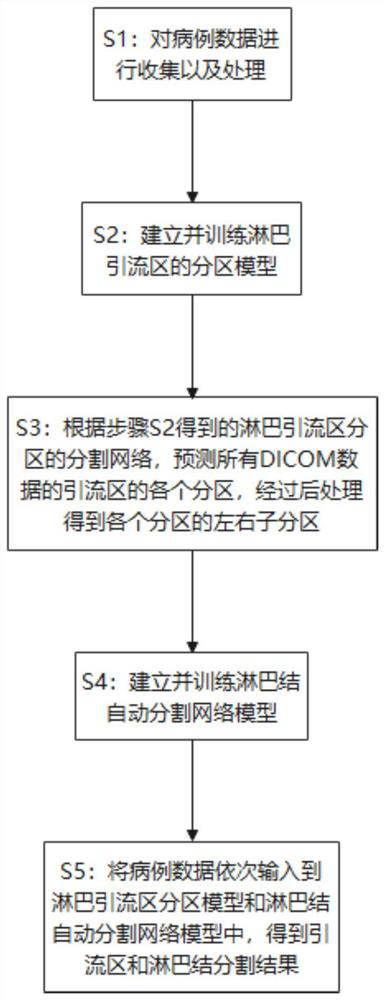
[0042] The present invention provides an automatic delineation method for the drainage area and metastatic lymph nodes of head and neck nasopharyngeal carcinoma. Firstly, each partition of the drainage area is segmented, and then the image range of the segmented lymph nodes is narrowed according to the prediction results of each partition, so that the network pays more attention to the drainage. Inside the area, reducing false positive areas. The overall process is as follows figure 1 As shown, it specifically includes the following steps:

[0043] S1: Collect and process case data.

[0044] The case data include DICOM images and the corresponding outline data of lymph nodes and drainage areas drawn by doctors.

[0045]Specifically, firstly, the DICOM images...


PUM


Abstract

The invention discloses an automatic delineation method for drainage areas and metastatic lymph nodes of head and neck nasopharyngeal carcinoma, and belongs to the field of medical image processing. The method comprises the following steps: S1, collecting and processing case data, wherein the case data comprises DICOM images; S2, establishing and training a lymphatic drainage area partition model; S3, predicting each partition of the drainage area of all DICOM data according to a partition network of a lymphatic drainage area partition obtained in the step S2, and performing post-processing to obtain left and right sub-partitions of each partition; S4, establishing and training a lymph node automatic segmentation network model; and S5, sequentially inputting the case data into the lymphatic drainage area partition model and the lymph node automatic segmentation network model to obtain a drainage area and lymph node segmentation result. According to the invention, the burden of a doctor for target delineation during radiotherapy can be reduced, the delineation efficiency of the doctor is improved, and the delineation subjectivity of the doctor is reduced.

Description

technical field [0001] The invention belongs to the field of medical image processing, and in particular relates to an automatic delineation method for head and neck nasopharyngeal carcinoma drainage area and metastatic lymph nodes based on deep learning. Background technique [0002] The lymphatic network is widely distributed in the nasopharyngeal region of the human head and neck, and nasopharyngeal carcinoma has a high rate of lymph node metastasis. Radiation therapy, as one of the most important treatment methods for nasopharyngeal carcinoma, requires doctors to accurately outline the gross tumor volume (GTV for short) and the corresponding subclinical target volume (Clinical target volume) on CT images. , referred to as CTV). Among them, GTV refers to the gross tumor area seen in imaging examination or clinical physical examination, specifically including the primary tumor (GTVp) and metastatic lymph nodes (GTVn). CTV includes subclinical lesions with a certain proba...

Claims


Application Information

Patent Timeline
Patent Type & Authority Applications(China)
IPC IPC(8): G16H30/40G16H30/20G06T7/13G06T7/187G06N3/04
CPCG16H30/40G16H30/20G06T7/13G06T7/187G06T2207/10081G06T2207/30096G06T2207/20081G06T2207/20104G06N3/045
Inventor 魏军蒋雪田孟秋谢培梁
Owner PERCEPTION VISION MEDICAL TECH CO LTD
Why Eureka
Try Eureka