
Fundus three-dimensional model establishment method, fundus camera, device and storage medium

A 3D model and establishment method technology, applied in the field of image processing, can solve the problems of large modeling error, limited shooting space, difficult 3D modeling of binocular cameras, etc., and achieve the effect of small modeling error

Active Publication Date: 2022-02-22
广东唯仁医疗科技有限公司
View PDF5 Cites 0 Cited by
  • Summary
  • Abstract
  • Description
  • Claims
  • Application Information

AI Technical Summary

Problems solved by technology

[0003] Since the fundus camera is used to capture the fundus through the pupil of the eyeball when taking fundus images, the shooting space is greatly limited, so it is difficult to use a binocular camera for 3D modeling
At present, in the related technology of using a monocular camera to model the three-dimensional model of the fundus, the eyeball is regarded as a standard ellipsoid, and then the fundus image on the two-dimensional plane obtained by shooting is projected onto the surface of the ellipsoid, thereby performing Fundus three-dimensional model modeling, but the eyeball is not a standard ellipsoid, and the shape of each person's eyeball is different, so the modeling error of this technology is very large

Method used


Image

  • Fundus three-dimensional model establishment method, fundus camera, device and storage medium
  • Fundus three-dimensional model establishment method, fundus camera, device and storage medium
  • Fundus three-dimensional model establishment method, fundus camera, device and storage medium

Examples


Embodiment Construction

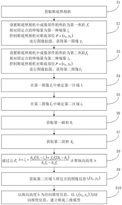
[0060] In this example, refer to figure 1 The method for establishing a three-dimensional fundus model includes the following steps:

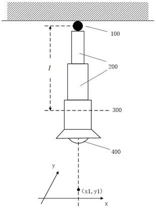
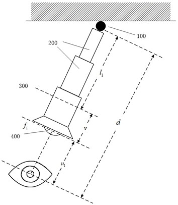
[0061] S1. Obtain a fundus camera; the fundus camera has a fixed point with a constant spatial position, and the imaging component of the fundus camera can be rotated and stretched relative to the fixed point;

[0062] S2. Set the first focal length of the imaging component in the fundus camera as f 1 , the first stretching amount relative to the fixed point is l 1 , control the fundus camera to the fundus position P= ( x 0 , y 0 ) for image capture to obtain the first image I 1 ;

[0063] S3. Set the second focal length of the imaging component in the fundus camera as f 2 , the second expansion relative to the fixed point is l 2 , control the fundus camera to the fundus position P= ( x 0 , y 0 ) for image capture to obtain the second image I 2 ;

[0064] S4. In the first image I 1 Determine the first region in i 1 ;...


PUM


Abstract

The invention discloses a method for establishing a three-dimensional model of the fundus, a fundus camera, a computer device and a storage medium. The method for establishing the three-dimensional model of the fundus includes taking two shots with different expansion and contraction amounts to obtain a first image and a second image, respectively in the first Determine the first area and the second area in the image and the second image, obtain the first area of ​​the first area, and the second area of ​​the mapping area corresponding to the second area in the first image, and use the zoom amount, focal length, and first The parameters such as area determine the radial dimension information, and the image information obtained by shooting is used as the tangential dimension information to establish a three-dimensional fundus model. In the present invention, the same part of the fundus is photographed twice with different object distances, and the object distance is calculated according to the shooting parameters, thereby obtaining radial dimension information and tangential dimension information to establish a three-dimensional fundus model. The present invention uses measured data to establish a three-dimensional model of the fundus, and the modeling error is small. The invention is widely used in the technical field of image processing.

Description

technical field [0001] The invention relates to the technical field of image processing, in particular to a fundus three-dimensional model building method, a fundus camera, a computer device and a storage medium. Background technique [0002] The inner wall of the eyeball can also be called the fundus. By taking images of the fundus, doctors can observe the condition of the fundus, so as to diagnose and treat diseases. The fundus image is a two-dimensional planar image, which lacks spatial distribution information and cannot provide doctors with comprehensive information about the fundus, thus limiting the accuracy of doctor's disease diagnosis and treatment. Therefore, a three-dimensional model of the fundus is created. Through three-dimensional fundus The model shows the image information of the parts on the fundus and its spatial distribution to the doctor, which is helpful for the doctor to diagnose and treat the disease. [0003] Since the fundus camera is used to capt...

Claims


Application Information

Patent Timeline
Patent Type & Authority Patents(China)
IPC IPC(8): G06T7/55G06T7/62
CPCG06T7/55G06T7/62G06T2207/10016G06T2207/30041
Inventor 秦嘉安林
Owner 广东唯仁医疗科技有限公司
Learn More