
Endoscopic examination image acquisition method and device

An image acquisition and image technology, applied in the field of medical image processing, can solve problems such as limiting the range of activities of physicians, reducing physician work efficiency, increasing manpower burden, etc., to achieve the effect of improving work efficiency, improving endoscopic examination efficiency, and reducing manpower burden

Active Publication Date: 2021-11-23
青岛美迪康数字工程有限公司
View PDF9 Cites 2 Cited by
  • Summary
  • Abstract
  • Description
  • Claims
  • Application Information

AI Technical Summary

Problems solved by technology

However, because the pedals are fixed at the designated location, the doctor's range of activities is limited
An additional manpower burden is also added by providing assistants to physicians
[0003] In addition, in actual use, there is a certain delay from when the doctor observes a suitable image to triggering the image acquisition, so the collected image is often not what the doctor needs, and the doctor often freezes the image before collecting the image (the inspection equipment must have a frozen image functions), or continuously collect multiple images of the same part, and then select appropriate images from them when writing a report, which also reduces the work efficiency of physicians
[0004] For the above-mentioned problems existing in the existing endoscopic image acquisition, the prior art does not provide an effective solution

Method used


Image

  • Endoscopic examination image acquisition method and device
  • Endoscopic examination image acquisition method and device
  • Endoscopic examination image acquisition method and device

Examples


Embodiment 1

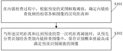
[0045] Embodiments of the present invention provide a method for collecting images of endoscopic examination, such as figure 1 As shown, the endoscopic image acquisition method includes:

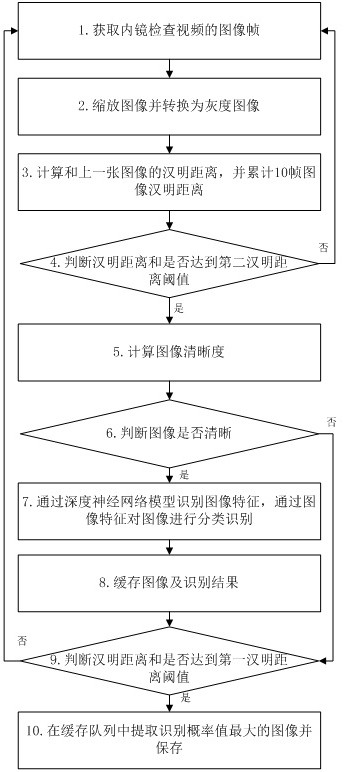
[0046] S101. During the endoscopic examination process, according to the preset image acquisition frame number threshold, determine the sum of the Hamming distances of the adjacent frame images of the endoscopic examination video; the video observed and displayed on the monitor of the endoscopy equipment;

[0047] S102. When the sum of the Hamming distances reaches a preset first Hamming distance threshold, save the image with the highest recognition probability or satisfying the preset recognition threshold from the pre-classified and recognized endoscopic examination images.

[0048] Wherein, the setting of the image acquisition frame number threshold can determine the accuracy of image acquisition, and optionally, the image acquisition frame number threshold is selected as 10 frames. In...

Embodiment 2

[0113] An embodiment of the present invention provides an endoscopic inspection device, and the endoscopic inspection includes: a memory, a processor, and a computer program stored in the memory and operable on the processor;

[0114] When the computer program is executed by the processor, the steps of the endoscopic image acquisition method according to any one of the first embodiment are realized.

Embodiment 3

[0116] An embodiment of the present invention provides a computer-readable storage medium, where an endoscopic image acquisition program is stored on the computer-readable storage medium, and when the endoscopic image acquisition program is executed by a processor, the implementation as in the first embodiment The steps of any one of the endoscopic image acquisition methods.

[0117] Embodiment 2 to Embodiment 3 may refer to Embodiment 1 during the specific implementation process, and have corresponding technical effects.


PUM


Abstract

The invention relates to an endoscopic examination image acquisition method and device. The endoscopic examination image acquisition method comprises the steps of determining the sum of Hamming distances between adjacent frames of images of an endoscopic examination video according to a preset image acquisition frame number threshold value in an endoscopic examination process; and when the sum of the Hamming distances reaches a preset first Hamming distance threshold value, storing an image with the highest recognition probability value or meeting a preset recognition threshold value from various types of endoscopic examination images which are classified and recognized in advance. According to the method, the endoscopic examination efficiency is improved, the working efficiency of a user is improved, and the manpower burden of endoscopic examination is reduced.

Description

technical field [0001] The invention relates to the field of medical image processing, in particular to an endoscopic image acquisition method and device. Background technique [0002] At present, physicians need to manually trigger the image acquisition function during the endoscopic examination. In actual work, the physician operates the instrument with both hands, and triggers the endoscopic image acquisition through the pedal or the equipped assistant. However, because the pedals are fixed at designated locations, the doctor's range of activities is limited. An additional human burden is also added through the assistants assigned to physicians. [0003] In addition, in actual use, there is a certain delay from when the doctor observes a suitable image to triggering the image acquisition, so the collected image is often not what the doctor needs, and the doctor often freezes the image before collecting the image (the inspection equipment must have a frozen image functi...

Claims


Application Information

Patent Timeline
Patent Type & Authority Applications(China)
IPC IPC(8): A61B1/00G06K9/00G06K9/62G06N3/04G06N3/08
CPCA61B1/00004G06N3/08G06N3/045G06F18/22G06F18/24
Inventor 冯健常培佳邵学军杨延成
Owner 青岛美迪康数字工程有限公司
Learn More