Reconstruction of soft tissue-bone immune repair environment periosteum-bone complex and preparation method
An immune repair and complex technology, applied in tissue regeneration, prosthetics, medical science, etc., can solve the problem of no periosteum-bone complex, achieve coordination of early immune regulation of osteogenesis-angiogenesis events, and promote bone repair , The effect of low cytotoxicity of the scaffold
- Summary
- Abstract
- Description
- Claims
- Application Information
AI Technical Summary
Problems solved by technology
Method used
Image
Examples
Embodiment 1
[0044] Example 1: Periosteum-bone complex aimed at reconstructing soft tissue-bone immune repair environment and its preparation method
[0045] (1) Take material
[0046] The femurs of healthy adult large white pigs were taken within 12 hours of slaughter, the metaphysis was separated, the femoral shaft was collected, the internal marrow cavity was removed, and the femoral shaft was divided into 10mm*8mm pieces;
[0047] (2) Preprocessing
[0048] Take a certain amount of flaky bone, place it on a low-temperature (4°C) shaker and shake at 100 rpm, and rinse it with PBS buffer containing protease inhibitors for 3 times for 10 min each time to remove blood, attached fat fragments and other impurities;
[0049] (3) Decalcification
[0050] The pretreated flaky bones were placed in a 20% (w / v) EDTA-2Na solution for ultrasonic decalcification for 12 days, the ultrasonic parameters were 40kHZ, and the temperature was 22°C;
[0051] (4) Obtaining the periosteum-bone complex
[0...
example 1
[0063] 1. In Example 1, the macroscopic view and decellularization assessment of the periosteum-bone complex scaffold
[0064] The general appearance and CT scan of the periosteum-bone complex scaffold, such as figure 1 .
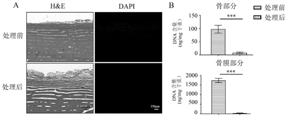
[0065] 2. In Example 1, Decellularization Assessment of Periosteum-Bone Complex Scaffolds
[0066] H&E staining showed that no cells and no nuclear components remained, and DNA quantitative detection almost did not contain DNA components, such as figure 2 .
[0067] 3. In Example 1, Microstructural Evaluation of Periosteum-Bone Complex Scaffolds
[0068] Scanning electron microscope was used to observe collagen arrangement, bone crystal structure and periosteum-bone interface connection structure of the present invention, such as image 3 .
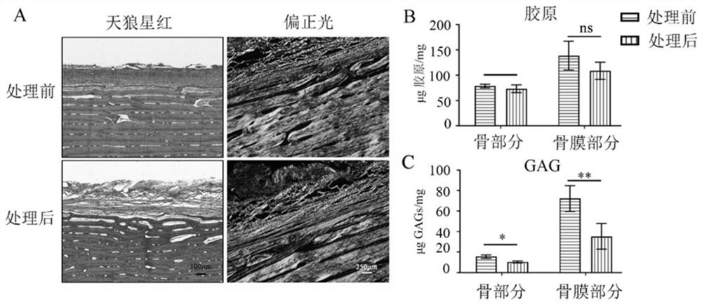
[0069] 4. Surface topography and mechanical evaluation of the periosteum-bone complex scaffold in Example 1
[0070] Atomic force microscope observes the microscopic surface morphology of the present invention, ...
Embodiment 2
[0081] Example 2: Periosteum-bone complex aimed at reconstructing soft tissue-bone immune repair environment and its preparation method
[0082] (1) Take material
[0083] The femurs of healthy adult large white pigs were taken within 12 hours of slaughter, the metaphysis was separated, the femoral shaft was collected, the internal marrow cavity was removed, and the femoral shaft was divided into 10mm*8mm pieces;
[0084] (2) Preprocessing
[0085] Take a certain amount of flaky bone, place it on a low-temperature (4°C) shaker and shake at 100 rpm, and rinse it with PBS buffer containing protease inhibitors for 3 times for 10 min each time to remove blood, attached fat fragments and other impurities;
[0086] (3) Decalcification
[0087] The pretreated flaky bones were placed in a 20% (w / v) EDTA-2Na solution for ultrasonic decalcification for 12 days, the ultrasonic parameters were 40kHZ, and the temperature was 22°C;
[0088] (4) Obtaining the periosteum-bone complex
[0...
PUM
 Abstract
Description
Claims
Application Information
 - R&D
- Intellectual Property
- Life Sciences
- Materials
- Tech Scout
- Unparalleled Data Quality
- Higher Quality Content
- 60% Fewer Hallucinations
Browse by: Latest US Patents, China's latest patents, Technical Efficacy Thesaurus, Application Domain, Technology Topic, Popular Technical Reports.
© 2025 PatSnap. All rights reserved.Legal|Privacy policy|Modern Slavery Act Transparency Statement|Sitemap|About US| Contact US: help@patsnap.com


