Magnetic resonance imaging method and device, computer equipment and storage medium

A technology of magnetic resonance imaging and computer programs, which is applied in the fields of magnetic resonance measurement, nuclear magnetic resonance image system for measurement, magnetic variable measurement, etc., can solve problems such as high difficulty in cooperation and long breath-holding time, and save scanning time and reduce The effect of holding breath time and increasing scanning efficiency

Pending Publication Date: 2022-04-12
SHANGHAI UNITED IMAGING HEALTHCARE
View PDF0 Cites 0 Cited by
  • Summary
  • Abstract
  • Description
  • Claims
  • Application Information

AI Technical Summary

Problems solved by technology

[0004] However, the MOLL technique requires a longer breath-holding time, and it is more difficult for the elderly and children to cooperate

Method used


Image

  • Magnetic resonance imaging method and device, computer equipment and storage medium
  • Magnetic resonance imaging method and device, computer equipment and storage medium
  • Magnetic resonance imaging method and device, computer equipment and storage medium

Examples


Embodiment Construction

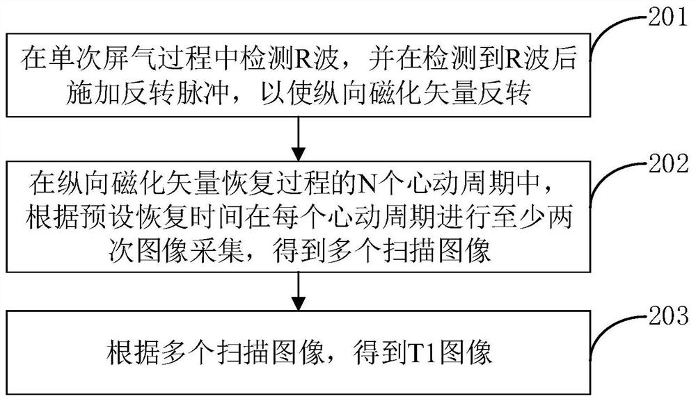
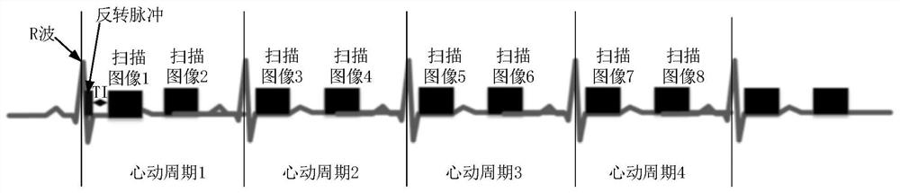
[0055] In order to make the purpose, technical solution and advantages of the present application clearer, the present application will be further described in detail below in conjunction with the accompanying drawings and embodiments. It should be understood that the specific embodiments described here are only used to explain the present application, and are not intended to limit the present application.

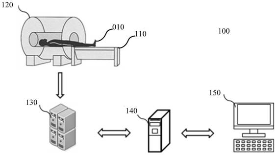
[0056]The magnetic resonance imaging method provided by the application can be applied to such as figure 1 shown in the application environment. The application environment is a magnetic resonance system. The magnetic resonance system 100 includes a bed body 110 , an MR scanner 120 and a processor 130 . The MR scanner 120 includes a magnet, a radio frequency transmitting coil, a gradient coil and a radio frequency receiving coil. The bed body 110 is used to carry the target object 010, the radio frequency transmitting coil is used to transmit radio frequency pulses to the...


PUM


Abstract

The invention relates to a magnetic resonance imaging method and device, computer equipment and a storage medium. The method comprises the following steps: detecting an R wave in a single breath-holding process, and applying a reversal pulse after the R wave is detected, so that a longitudinal magnetization vector is reversed; in N cardiac cycles of the longitudinal magnetization vector recovery process, performing at least two times of image acquisition in each cardiac cycle according to preset recovery time to obtain a plurality of scanning images; wherein N is a positive integer; and obtaining a T1 image according to the plurality of scanning images. By adopting the method, the breath-holding time of the scanned object can be reduced.

Description

technical field [0001] The present application relates to the technical field of magnetic resonance imaging, in particular to a magnetic resonance imaging method, device, computer equipment and storage medium. Background technique [0002] Cardiac Magnetic Resonance Imaging (CMRI) has good tissue contrast and is non-invasive and harmless to the human body. It has been widely used in the evaluation of the shape, function and tissue characteristics of the heart in recent years. Myocardial longitudinal relaxation time will change accordingly when the myocardial water content and extracellular environment change. The longitudinal relaxation time quantitative imaging (T1 mapping) technology can quantitatively reflect the tissue characteristics of the myocardium. The diffuse lesions such as myocardial edema, fibrosis, fatty infiltration and iron deposition were detected by quantitative measurement of the initial T1 value and the T1 value after injection of contrast medium. [000...

Claims


Application Information

Patent Timeline
Patent Type & Authority Applications(China)
IPC IPC(8): A61B5/055G01R33/48
Inventor 赵乐乐
Owner SHANGHAI UNITED IMAGING HEALTHCARE
Why Eureka
Try Eureka